ACEP ID:

Left Arrow Right Arrow Down Arrow Up Arrow Left Arrow Right Arrow Down Arrow Up Arrow
Section IconUltrasound-Guided Bicipital Tendinitis Injection
Overview
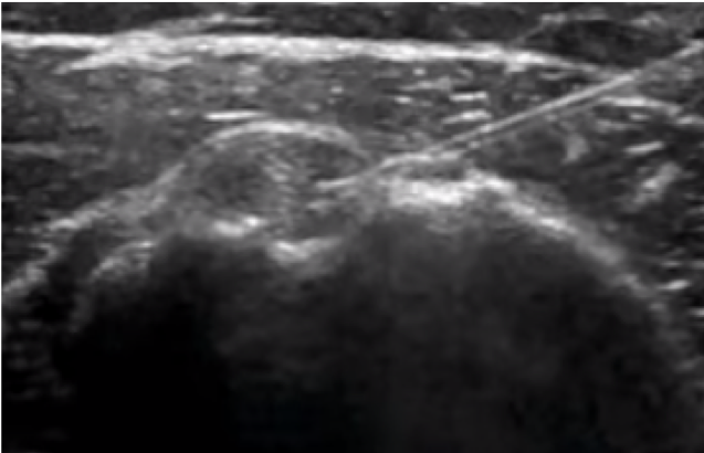
Bicipital tendinitis is a condition caused by inflammation of the long head of the biceps muscle’s tendon. It’s commonly caused by sudden overuse of the muscle and rotator cuff pathology such as muscle or tendon tears, chronic impingement, multidirectional shoulder instability, calcifications, and deconditioning. Signs in physical examination correlating with bicipital tendinitis are pain at the bicipital groove and a positive provocative test such as Speed’s test and Yergason’s test, although studies have shown that their sensitivity might be poor.1
US may assist in making the diagnosis. In Figure 1 a normal biceps tendon can be appreciated at the bicipital groove. In Figure 2 there’s synovial thickening with an effusion surrounding the long head biceps muscle’s tendon.
Indications
Tenderness at bicipital groove
Positive Yergason’s test: have the patient in seating position with the affected arm resting at his side, elbow flexed at 90o, and forearm in pronation. Grab the patient’s forearm at the wrist to resist supination, and direct the patient to supinate his forearm against your resistance. Pain reflected in the bicipital groove indicates a positive test. Figure 3
Positive Speed’s test: have the patient extend the affected arm with the elbow extended and wrist supinated. Grab the patient’s forearm and direct the patient to forward flex her shoulder against your resistance. Pain reflected in the bicipital groove indicates a positive test. Figure 4
Ultrasonographic evidence of effusion surrounding the long head biceps muscle’s tendon.
Contraindications
Cellulitis at the bicipital groove or area of injection
Allergy to anesthetic
Equipment & Dosing
US machine with a high-frequency linear probe
Sterile gloves
Alcohol swabs or chlorhexidine
10-mL syringe
21- or 23-gauge 1.5-in needle
Adhesive bandage
Steroid: choose one of the following:
  • Triamcinolone acetonide (Kenalog): 0.5 mL of 20 mg/mL (10 mg)
  • Methylprednisolone (Depo-Medrol): 0.25 mL of 40 mg/mL (10 mg)
  • Betamethasone sodium phosphate/betamethasone acetate (Celestone Soluspan): 0.5 mL of 6 mg/mL (3 mg)
Lidocaine 1% or 2% or Bupivacaine 0.25% or 0.5%: 1 to 2 mL
Procedure & Administration
Obtain consent
Patient should be seated comfortably with elbow slightly flexed and forearm supinated.
Place the high-frequency linear US probe at the bicipital groove with the probe marker pointing to patient’s right side. Scan the area at the point of maximum tenderness to find the appropriate injection site. Once you locate the appropriate injection site, mark the injection site. Figure 5
Prepare the patient using aseptic technique. Sterilize a large area surrounding the injection site with alcohol swabs or chlorhexidine.
Place the high-frequency linear US probe at the previously marked area in the bicipital groove. Place small wheal of local anesthetic then advance needle using an in-plane approach (Figure 6). Figure 6
Advance needle until it reaches the tendon sheath (Figure 7). Aspirate to make sure you are not in any vessel then slowly inject the contents of the syringe.
There should be no significant resistance. If you feel resistance, it would indicate that the needle has pierced the tendon. If so, slowly retract the needle until there’s no more resistance. On the US screen, correct injection of the tendon sheath will show distention around the tendon as the medication mixture is injected.
Remove the needle, apply pressure with gauze or dressing, and place a bandage at the injection site.
Perform a physical examination to look for changes in pain score, strength, range of motion, and provocative tests . Figure 3 - Figure 4
Perform neurologic examination to ensure no neurologic deficit is present.
Complications
Bicipital tendon rupture with intratendinous injection
Infection
Trauma to bicipital tendon (Damaged tendons might be more prone to rupture.)
Charting & Documentation
Document:
  • Performance of neurologic examination before and after the procedure, no neurologic deficit present
  • Any changes noted on physical examination after the procedure
  • Use of aseptic technique during the procedure
Discharge Procedure
Instruct the patient to:
  • Avoid overhead activities, vigorous exercise, and lifting heavy objects in the first 48 to 72 hours while tendon heals.
  • Take NSAIDs to reduce pain and inflammation.
  • Apply ice to the area 15 to 20 minutes 2 or 3 times a day for several days to decrease inflammation.
  • Begin gentle stretching of the scapula and rotator cuff muscles 3 to 5 days after the procedure.
  • Start exercising after shoulder pain stops.
  • Follow up with the primary care physician to assess pain resolution or to examine other causes if pain persists, as bicipital tendinitis is usually associated with other pathologies of the shoulder.
References
  1. Chen HS, Lin SH, Hsu YH, et al. A comparison of physical examinations with musculoskeletal ultrasound in the diagnosis of biceps long head tendinitis. Ultrasound Med Biol. 2011 Sep;37(9):1392–8.
  2. Hashiuchi T, Sakurai G, Morimoto M, et al. Accuracy of the biceps tendon sheath injection: ultrasound-guided or unguided injection? A randomized controlled trial. J Shoulder Elbow Surg. 2011 Oct;20(7):1069–73.
  3. Churgay CA. Diagnosis and treatment of biceps tendinitis and tendinosis. Am Fam Physician. 2009 Sep 1;80(5):470–6.
  4. Krabak BJ. Biceps tendinopathy. In: Frontera WR, Silver JK, Rizzo TD, eds. Essentials of Physical Medicine and Rehabilitation: Musculoskeletal Disorders, Pain, and Rehabilitation. 3rd ed. Philadelphia, PA:Saunders;2015:58–61.
  5. Waldman SD. Bicipital tendon injection. In: Waldman SD, ed. Atlas of Pain Management Injection Techniques. 3rd ed. Philadelphia, PA:Saunders;2013:83–6.
  Back To Top

Figure 1

Normal biceps tendon in the bicipital

Figure Image 1

Close

Figure 2

Synovial hypertrophy and surrounding groove between the greater and lesser tubercles effusion of an inflamed long head biceps of the humerus muscle’s tendon

Figure Image 2

Close

Figure 3

Yergason’s test

Figure Image 3

Close

Figure 4

Speed’s test

Figure Image 4

Close

Figure 5

High-frequency US probe placed at bicipital groove showing the long head biceps muscle’s tendon at the center of the screen

Figure Image 5

Close

Figure 6

High-frequency US probe placed at bicipital groove showing the long head biceps muscle’s tendon at the center of the screen

Figure Image 6

Close

Figure 7

US image of the steroid injection using an in-plane approach (Notice the needle at the upper right corner entering the tendon sheath.)

Figure Image 7

Close
[ Feedback → ]