ACEP ID:

Left Arrow Right Arrow Down Arrow Up Arrow Left Arrow Right Arrow Down Arrow Up Arrow
Section IconIntra-Articular Posterior Shoulder Injection
Overview
Intra-articular posterior shoulder injections are as effective as intravenous sedation for treating pain associated with shoulder dislocation.1,2
Ultrasound guidance increases the accuracy of shoulder injections compared to landmark-based techniques.3
Ultrasound can also confirm dislocation and reduction of the humeral head when landmarks are difficult to palpate.
The anterior technique can be used, but the posterior technique is easier and more effective.4
Indications
Reduction of a dislocated shoulder
Contraindications
Allergy to anesthetic
Altered or unconscious patient
Infection overlying the area
Neurologic deficit in the affected extremity
Equipment & Dosing
Ultrasound machine with a high-frequency linear probe
  • Low-frequency curvilinear (1-5MHz) probe might be required for muscular and obese patients
Sterile ultrasound probe cover and gel
Sterile gloves
Skin disinfectant and antiseptic
Bupivacaine or ropivacaine 0.25-0.5% or lidocaine 1-2%
  • Always calculate maximum dose, bupivacaine and ropivacaine 3 mg/kg and lidocaine 5 mg/kg
20- to 30-mL syringe
18-gauge needle
Intravenous access
Anatomy & Innervation
The shoulder is a ball-and-socket joint composed of the humeral head and the glenoid fossa, which arises from the scapula and is lined by a fibrocartilaginous layer, the glenoid labrum.
Anterior shoulder dislocations (common) occur when an external force causes abduction, extension, and external rotation of the humeral head, pushing it inferiorly below the glenoid fossa.
  • The pectoralis and biceps muscles subsequently contract and pull the humerus anteriorly to a location just below the glenoid fossa or coracoid.
  • The axillary and musculocutaneous nerves are at risk for injury.
Posterior shoulder dislocations (rare) occur when there is forceful internal rotation and adduction of the shoulder.
Procedure & Administration
Have the patient sit facing away from you, with the affected arm at the side in a neutral position. Figure 1
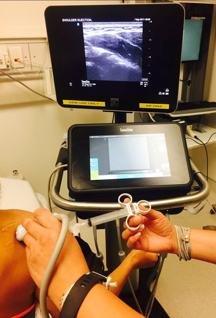
Place the ultrasound machine in front of the patient on the affected side.
Disinfect the skin.
Place a sterile cover over the ultrasound probe and apply sterile gel.
Place the probe in a transverse orientation over the infraspinatus and posterior glenohumeral joint, along the axis of the spine of the scapula and just caudal to the acromion.
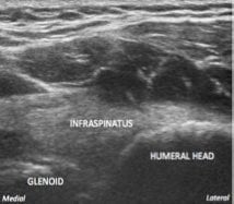
 Figure 2
Identify the humeral head, glenoid labrum, infraspinatus tendon, and joint capsule.
  • Normally, the humeral head is located in the glenoid fossa. Figure 3
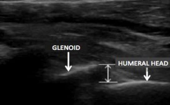
  • In an anterior shoulder dislocation, the humeral head is located in the far field (deep to the probe). Figure 4
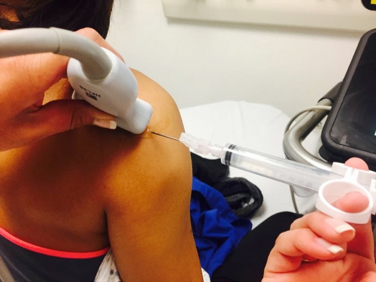
Place a skin wheal with local anesthetic lateral to the probe with a small-bore needle.
Enter the skin 1 cm lateral to the probe and advance the needle from the lateral to medial direction.
Keep the probe in a transverse orientation and use an in-plane technique.
Enter the shoulder joint between the posterior glenoid labrum and the insertion of the infraspinatus tendon on the hyperechoic (bright) humeral head.
Once in the joint, aspirate to make sure the needle is not in a vessel. It is common to get a flash of blood when you’re in the joint. 8
Inject 15-20 mL of local anesthetic into the joint space, visualizing the needle at all times. If it cannot be visualized, stop the procedure.
The patient should start to experience pain relief within 10 minutes. The duration of the block should be 3 to 8 hours, depending on the anesthetic used.
Complications
Accidental puncture or injection into the suprascapular or the circumflex scapular neurovascular structures
Infection
Local anesthetic systemic toxicity
  • Always calculate maximum dose (bupivacaine and ropivacaine 3 mg/kg and lidocaine 5 mg/kg).
Charting & Documentation
An ultrasound-guided shoulder injection requires a procedure note in the medical record.
Document:
  • Clinical examination before the procedure, including neurologic and vascular examinations of the upper extremity
  • Sterile preparation and local antiseptic used
  • Needle approach and confirmation of needle placement
  • Type and dose of anesthetic injected
  • Clinical examination immediately after the procedure
  • Complications
Special Considerations
Anticoagulation is not a contraindication, but care must be taken to avoid puncturing a deep vessel.
Discharge Procedure
Tell the patient about driving restrictions, work limitations, participation in sports, and so on as appropriate.
Where to Learn More
References

Watch the reference video

  Back To Top

Figure 1

Intra Articular Posterior Shoulder

Position the patient facing away, with the affected extremity in a neutral position.

Figure 1 Image

Close

Figure 2

Intra Articular Posterior Shoulder

Hold the probe in a transverse orientation over the infraspinatus and posterior glenohumeral joint, just caudal to the acromion. Enter the skin lateral to the probe and advance from the lateral to medial direction.

Figure 2 Image

Close

Figure 3

Intra Articular Posterior Shoulder

Intact shoulder.

Figure 3 Image

Close

Figure 4

Intra Articular Posterior Shoulder

Anteriorly dislocated shoulder.

Close
[ Feedback → ]