ACEP ID:

Left Arrow Right Arrow Down Arrow Up Arrow Left Arrow Right Arrow Down Arrow Up Arrow
Section IconOccipital Nerve Blocks
Overview
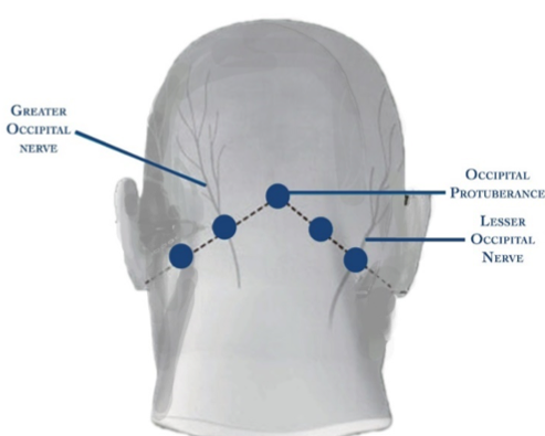
The greater and lesser occipital nerves originate from the neck and provide sensory input to either side of the scalp. They lack motor function. Occipital nerve blocks involve the injection of local anesthetic, with or without steroids, to inhibit the activity of these nerves in the following evidence-supported conditions. Figure 1
Indications
Occipital neuralgia
Cervicogenic headache
Cluster headache
Postconcussive headache
Postdural puncture headache
Medication overuse headache
Migraine
Contraindications
Amide/Ester allergy
Overlying cellulitis
Significant coagulopathy
Trauma or other landmark distortion
Equipment & Dosing
Chlorhexidine swabs or alcohol pads
Local anesthetic
22- to 25-gauge needle
10-cc syringe
Sterile gloves
Procedure & Administration
Palpate the occipital protuberance.
Locate the GON 2 cm lateral and 2 cm inferior to the protuberance.
Alternatively, draw an imaginary line between the occipital protuberance and the mastoid process. Locate landmarks at both 1/3 and 2/3 the distance along this line to target both the GON and LON, respectively. Figure 2
You may also palpate the occipital artery along the midpoint of this line and find the GON just medial.
Clean the area with an alcohol swab/chlorhexidine.
Insert needle until periosteum resistance is met, and retract slightly.
Aspirate to ensure you are not in a vessel.
Inject 2 to 3 cc of local anesthetic
Withdraw the needle. Then compress and massage the injection site to both stop bleeding and help diffuse anesthetic into tissue.
Dress the injection site.
Observe patient 15 minutes for adverse reactions.
Complications
Anesthetic allergy/anaphylaxis
Injection site reaction
Localized infection
Occipital arterial hematoma
Superficial bleeding
Transient lightheadedness/syncope
No risk of CSF leak, spinal cord injury, or pneumothorax
Special Considerations
Steroids (methylprednisolone 40 mg, dexamethasone 4 mg) are thought to prolong the duration of analgesic effect by reducing the influx of inflammatory cytokines and prostaglandin formation. Frequent use, however, is associated with risk of tissue necrosis, muscle wasting, and tendon rupture.
Ultrasonography can be used to provide targeted therapy and reduce required anesthetic volume. (See following videos.)
Devices (OcciGuide) improve landmark accuracy. Figure 3
References
  1. Landy S, Rice K, Lobo B. Central sensitization and cutaneous allodynia in migraine: implications for treatment. CNS Drugs. 2004;18(6):337–42.
  2. Dubin AE, Patapoutian A. Nociceptors: the sensors of the pain pathway. J Clin Invest. 2010 Nov;120(11):3760–72.
  3. National Institute of Neurological Disorders and Stroke. Occipital neuralgia. 2017.
  Back To Top

Figure 1

Figure Image 1

Close

Figure 2

Figure Image 2

Close

Figure 3

Devices (OcciGuide) improve landmark accuracy

Figure Image 3

Close
[ Feedback → ]