ACEP ID:

Left Arrow Right Arrow Down Arrow Up Arrow Left Arrow Right Arrow Down Arrow Up Arrow
Section Icon Posterior Tibial Nerve Block
Overview
Posterior tibial nerve (PTN) blocks provide analgesia to the calcaneus and the majority of the plantar surface of the foot, areas that are difficult to anesthetize with local infiltration.1-4
PTN blocks alone don’t provide adequate anesthesia for ankle fractures but can be used with other regional blocks.
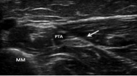
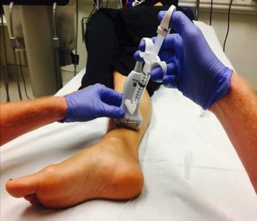
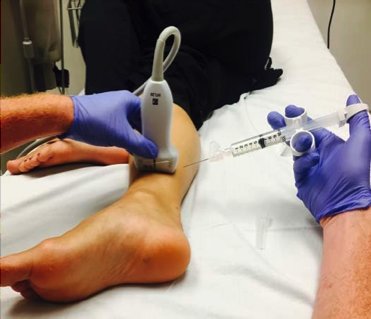
PTN blocks can be performed using landmark-based or ultrasound-guided techniques. The use of ultrasound has been shown to improve the success of sensory block.5
Indications
Pain associated with calcaneal fractures and foot lacerations, burns, infections, and foreign body removal
Contraindications
Allergy to local anesthetic
Altered or unconscious patient
Infection overlying the area to be blocked
Neurologic deficit in the affected extremity
Compartment syndrome of the foot occurs in 10% patients with calcaneal fractures and can be a contraindication for a PTN block due to its effect on the clinical examination.6 Discuss this with the orthopedist or podiatrist before performing the block.
Equipment & Dosing
Ultrasound machine with a high-frequency linear probe
Sterile ultrasound probe cover and gel
Sterile gloves
Skin disinfectant and antiseptic
Bupivacaine or ropivacaine 0.25-0.5% or lidocaine 1-2%
  • Always calculate maximum dose, bupivacaine and ropivacaine 3 mg/kg and lidocaine 5 mg/kg
Cardiac monitor
10- to 20-mL syringe
Intravenous access
20- to 30-gauge 3.5 inch noncutting spinal needle (reduces risk of nerve or vascular injury)
Anatomy & Innervation
The sciatic nerve bifurcates into the posterior tibial and common peroneal nerves at the popliteal fossa.
The posterior tibial nerve travels with the posterior tibial artery in a neurovascular bundle between the superficial and deep compartments of the lower leg.
This bundle passes under the flexor retinaculum and posterior to the medial malleolus in the following order (anterior to posterior): tibialis posterior tendon, flexor digitorum tendon, posterior tibial artery, posterior tibial nerve, flexor hallucis longus tendon.
The posterior tibial nerve is a mixed motor and sensory nerve. It provides motor innervation to the flexor muscles of the ankle and foot and sensory innervation to parts of the heel and the majority of the sole of the foot.
Procedure & Administration
Positioning
Have the patient lie supine with the affected lower extremity externally rotated at the hip and flexed at the knee.
Elevate the affected foot.
Have the patient lie on the affected side with the medial ankle exposed and the unaffected lower extremity held out of the way.
Localizing the nerve
Disinfect the skin proximal and posterior to the medial malleolus.
Place a sterile cover over the ultrasound probe and apply sterile gel.
Hold the high-frequency linear ultrasound probe in a transverse orientation to the long axis of the leg, approximately 3 cm proximal to the medial malleolus.
Slide the probe proximally to identify the following structures (anterior to posterior): Figure 1
  • Medial malleolus (hyperechoic)
  • Posterior tibial artery (hypoechoic and pulsatile)
  • Posterior tibial nerve (hyperechoic and approximately 3 mm in diameter)
Performing the nerve block
Out-of-plane technique: often easier to perform than the in-plane technique. Figure 2
  • Place a local skin wheal of anesthetic just adjacent to the middle of the probe.
  • With a spinal needle attached to the anesthetic syringe, enter the skin at the middle of the probe at a steep angle. The needle tip should be visualized at all times.
In-plane technique: Figure 3
  • Place a local skin wheal of anesthetic just adjacent to the probe in transverse orientation.
  • With a spinal needle attached to the anesthetic syringe, enter the skin from the posterior direction, 1 cm adjacent to the probe. Entry of the needle should generally be at a 45-degree angle. The needle should be visualized at all times.
Slowly inject 2 to 3 mL of anesthetic to confirm location. Anesthetic will have a hypoechoic (dark) appearance. If patient feels a sharp electric type pain with injection remove needle slightly and re-inject. Patient should feel pressure but not electric shooting pain.
Using V-shaped redirections of the needle, inject 5 to 8 mL of anesthetic around the nerve. The needle should not penetrate the nerve at any point.
  • Full circumferential spread of anesthetic around the nerve is ideal but not required.
If the needle cannot be visualized, stop the procedure.
The patient should start to experience pain relief within 15 minutes and full blockade after 30 minutes. The duration of the block is 3 to 8 hours, depending on the anesthetic used.
Complications
Infection
Intraneural injection
Local anesthetic systemic toxicity
  • Always calculate maximum dose (bupivacaine and ropivacaine 3 mg/kg and lidocaine 5 mg/kg).
Vascular puncture and hematoma
Charting & Documentation
A posterior tibial nerve block requires a procedure note in the medical record.
Document:
  • Clinical examination before the procedure, including neurologic and vascular examinations of the extremity
  • Sterile preparation and local antiseptic used
  • Needle approach and confirmation of needle placement
  • Type and dose of anesthetic injected
  • Clinical examination immediately after the procedure
  • Complications
Special Considerations
Anticoagulation is not a contraindication, but care must be taken to avoid puncturing a blood vessel.
Discharge Procedure
The patient will be admitted, transferred, or discharged depending on the injury.
Tell the patient to be discharged that some activities, such as walking and driving, might be difficult.
References
  1. Herring A. Ultrasound-guided posterior tibial nerve blocks. Highland EM Ultrasound Fueled pain management website. Accessed June 22, 2018.
  2. Myerson MS, Ruland CM, Allon SM. Regional anesthesia for foot and ankle surgery. Foot Ankle. 1992;13(5):282-288.
  3. Clattenburg E, Herring A, Hagn C, et al. ED ultrasound-guided posterior tibial nerve blocks for calcaneal fracture analgesia. Am J Emerg Med. 2016;34(6):1183.e1-e3.
  4. Soares LG, Brull R, Chan VW. Teaching an old block a new trick: ultrasound-guided posterior tibial nerve block. Acta Anaesthesiol Scand. 2008 ;52(3):446-447.
  5. Redborg KE, Antonakakis JG, Beach ML, et al. Ultrasound improves the success rate of a tibial nerve block at the ankle. Reg Anesth Pain Med. 2009;34: 256-260.
  6. Kalsi R, Dempsey A, Bunney EB. Compartment syndrome of the foot after calcaneal fracture. J Emerg Med. 2012;43:e101-e106.

Watch the reference video

  Back To Top

Figure 1

Posterior Tibial Nerve Block

On ultrasound, the posterior tibial nerve (arrow) appears hyperechoic (bright) and is located posterior to the hypoechoic (dark) posterior tibial artery (PTA). The medial malleolus appears as a hyperechoic (bright) line with shadowing.

Figure 1 Image

Close

Figure 2

Posterior Tibial Nerve Block

Out-of-plane technique.

Figure 2 Image

Close

Figure 3

Posterior Tibial Nerve Block

In-plane technique.

Figure 3 Image

Close
[ Feedback → ]