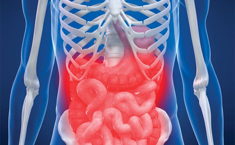
Midriff Mayhem: Abdominal Compartment Syndrome in the ICU
Objectives
- To understand how abdominal compartment syndrome (ACS) contributes to the morbidity and mortality of our patients
- To discuss a standardized approach to the diagnosis of ACS
- To review the medical management of intra-abdominal hypertension (IAH) and ACS

As your critically ill patient takes a turn for the worst with worsening hypotension, encephalopathy, and diminishing urine output, it is important to consider abdominal compartment syndrome (ACS) in your differential diagnosis. Intra-abdominal hypertension (IAH) and abdominal compartment syndrome (ACS) are conditions that exist on a spectrum. ACS is more commonly considered in the surgical population, as it is a well-known potential post-operative complication. Nevertheless, IAH and ACS can exist and will carry a high degree of morbidity and mortality in both surgical and medical ICU patients.1 High risk patients should be screened for IAH, so that we can proactively take measures to mitigate the risk of progression to ACS. When ACS is suspected, surgical consultation should be made early on, in order to reduce the morbidity and mortality associated with this potentially fatal condition.
A normal intra-abdominal pressure is less than 12 mmHg. There are four stages of IAH, which range from an abdominal pressure of 12-15 mmHg (Grade 1) to an abdominal pressure over 25 mmHg (Grade 4). An increasing grade of IAH has been associated with worsening morbidity and mortality.3 ACS is diagnosed when intra-abdominal pressure is greater than 20 mmHg (IAH Grade 3 or 4) in the setting of new organ dysfunction.
In a 2018 study, the incidence of IAH was reported to be approximately 34% on admission to a mixed medical, surgical and trauma ICU population.1 On admission, the incidence of ACS was 4%, while an additional 6% of patients developed ACS during their ICU stay. We know that severe IAH, and certainly ACS, can lead to an array of physiologic consequences, which may lead to multi-system organ dysfunction. The first sign of organ dysfunction is usually a drop in urine output. As the renal parenchymal pressure rises, venous drainage is impaired, leading to renal venous congestion with a subsequent drop in GFR and oliguria. In patients requiring mechanical ventilation, you may note an increase in airway pressures on the ventilator as the diaphragm is pushed superiorly. This pressure on the diaphragm will directly compress the lungs, resulting in both hypoxemic and hypercapnic respiratory failure. Similarly, external compression of the inferior vena cava can drastically reduce venous return and therefore cardiac output, presenting similarly to obstructive shock. In the gastrointestinal tract, mesenteric blood flow becomes severely diminished, leading to bowel ischemia and edema, which enters a cycle of worsening abdominal pressure as the bowel swells and can also increase the risk of bacterial translocation. Furthermore, the elevated abdominal pressures can directly press on the lumbar venous plexus, prohibiting the drainage of cerebrospinal fluid and leading to elevated intracranial pressure. In critical care medicine, we know that with each organ systems that fails, our patients’ mortality rises further. By contributing to multi-system organ dysfunction, IAH may be insidiously working against the recovery of some of our most critically ill patients and, at its most severe, can develop into abdominal compartment syndrome.
ICU patients are the highest risk patients for developing IAH and ACS, just by the nature of being critically ill. Specific risk factors for IAH/ACS include anything that will lead to one of the following:
- Reduced abdominal wall compliance (ie, obesity, circumferential burns, history of skin removal surgery)
- Increased intra-luminal contents (ie, ileus, bowel obstruction)
- Increased extra-luminal / intra-abdominal contents (ie, ascites, tumor, fluid collections)
- Third spacing (ie, severe pancreatitis, massive fluid resuscitation, sepsis)
If your patient is critically ill and has at least one risk factor for developing IAH, the World Society of Abdominal Compartment Syndrome (WSACS) recommends screening for elevated intra-abdominal pressure as frequently as every four-six hours, or at least once daily.2
The diagnosis of ACS rests on the measurement of intra-abdominal pressure in a patient with new organ dysfunction. Physical exam can aid your suspicion for the diagnosis but cannot be used to rule in or rule out the disease.4 The gold standard for estimating intra-abdominal pressure is via the measurement of intra-vesicular, or bladder, pressures. This is best done with the patient in a supine, generally relaxed position and measured at end-expiration. The pressure transducer should be along the mid-axillary line at the level of the iliac crest. When instilling fluid to create the continuous column of fluid from the bladder to the pressure transducer, it’s important to limit the volume of fluid to less than 25 milliliters in order to avoid falsely elevating the bladder pressure. For the most accurate measurement, consider neuromuscular blockade if your patient is already intubated.
The treatment for ACS is a decompressive laparotomy. Those who require this aggressive procedure will already carry a high degree of morbidity and mortality. Early surgical consultation is prudent, as the best outcomes occur following prompt diagnosis and intervention, ideally within 24 hours of the onset of ACS. Some laparotomies may be avoidable with aggressive medical management of IAH to address each of the above risk factors. Adequate pain control and promotion of relaxation will help avoid additional increases in abdominal pressure. Neuromuscular blockade will reduce intra-abdominal pressures and has been shown to improve organ function, although there is no proven mortality benefit with this technique.5 In the setting of worsening IAH, tube feeds should be held, and in the absence of bowel obstruction, pro-kinetic agents should be used. Additionally, attempts should be made at both nasogastric and rectal decompression. Drainage of intra-abdominal fluid, such as ascites or large fluid collections, may also be helpful. Finally, we should strive to avoid excessive IV fluids and correct positive fluid balances, when possible.
In conclusion, ACS carries a high degree of morbidity and mortality and must be on the differential diagnosis for all critically ill patients. Prompt diagnosis and treatment are needed to optimize outcomes. Screening of high-risk patients by measuring bladder pressure to estimate intra-abdominal pressure is a safe and easy way to detect IAH and ACS at an earlier stage. In those found to have IAH, there are a variety of noninvasive management strategies recommended by the WSACS.2
References
- Murphy PB, Parry NG, Sela N, et al. (2018). Intra-abdominal hypertension is more common than previously thought: A prospective study in a mixed medical-surgical ICU. Crit Care Med. 2018;46(6):958–64. https://doi.org/10.1097/CCM.0000000000003122
- Kirkpatrick AW, Roberts DJ, De Waele J, et al. Intra-abdominal hypertension and the abdominal compartment syndrome: Updated consensus definitions and clinical practice guidelines from the World Society of the Abdominal Compartment Syndrome. Intensive Care Med. 2013;39(7):1190–1206. https://doi.org/10.1007/s00134-013-2906-z
- Reintam Blaser A, Regli A, De Keulenaer B, et al. Incidence, risk factors, and outcomes of intra-abdominal hypertension in critically ill patients - a prospective multicenter study (IROI Study). Crit Care Med. 2019 Apr;47(4):535-42. doi: 10.1097/CCM.0000000000003623. PMID: 30608280; PMCID: PMC6426342.
- Sugrue M, Bauman A, Jones F, et al. Clinical examination is an inaccurate predictor of intraabdominal pressure. World J. Surg. 2002 Dec;26(12):1428-31. https://doi.org/10.1007/s00268-002-6411-8
- De Waele JJ, Benoit D, Hoste E, et al. A role for muscle relaxation in patients with abdominal compartment syndrome? Intensive Care Med. 2003:29(2):332. https://doi.org/10.1007/s00134-002-1578
Julieta Lacey, MD