Coral Snake Envenomations: Just Keep Breathing
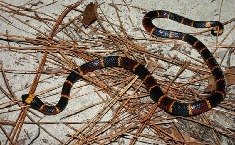
Image Above: “Coral snake” by MyFWC Florida Fish and Wildlife is licensed under CC BY-ND 2.0
Patient Case
A 63-year-old male presented to the emergency department via EMS 1 hour after sustaining a bite from the eastern coral snake, Micrurus fulvius. The patient had found the snake in his backyard, and he sustained the bite to his right middle finger while handling the snake in an attempt to remove it from his yard. On arrival to the ED, the patient reported mild swelling to the distal right middle finger and pain in the finger radiating to his shoulder along with the sensation of muscle stiffness and tingling in the hand.
On arrival, his vital signs were a temperature of 37 degrees Celsius, heart rate of 80 beats per minute, blood pressure of 165/95 mm Hg, respiratory rate of 16 breaths per minute, and oxygen saturation of 99% on room air. His physical examination was notable for a superficial bite mark with a small amount of dried blood on the flexor surface of the right middle finger distal phalynx with associated mild swelling and a capillary refill of less than 2 seconds. His right hand demonstrated full range of motion and 5/5 strength in the bilateral fingers, wrists, elbows, and shoulders. The remainder of his neurologic examination was unremarkable, with cranial nerves II-XII intact, normal sensation, normal muscle strength and tone in the lower extremities, normal reflexes, and normal coordination. He did not demonstrate any bulbar findings. His cardiac, pulmonary, and abdominal examinations did not demonstrate any abnormal findings.
His laboratory workup included CBC, CMP, CPK, and PT/INR. No acute laboratory abnormalities were noted. His EKG showed normal rate and rhythm with normal intervals and no QRS widening. His negative inspiratory force (NIF) was found to be -70 cmH2O, and his forced vital capacity (FVC) was 5 L. An x-ray of the right hand was obtained which did not demonstrate any retained teeth or other foreign bodies. He received a tetanus shot.
The regional poison control center was consulted, and the toxicologist advised the patient to hold his left upper extremity at or above the level of the heart. The toxicologist suspected the patient sensation of muscle cramping and tingling was due to envenomation. At the recommendation of the toxicologist, 5 vials of North American coral snake antivenin (NACSA) (produced by Wyeth, a subsidiary of Pfizer) was transported from a local venom unit to the hospital. The risks and benefits of NACSA were explained to the patient, with benefits including the potential to avoid intubation and prolonged hospital course and risks including allergic reaction and serum sickness. The patient verbalized understanding and consented to receive NACSA, and his RN administered the dose of 5 vials.
The patient was admitted to the ICU for frequent neurologic checks and respiratory function monitoring. The patient’s vital signs remained stable over a period of 24 hours, and the patient did not develop a decline in neurologic or pulmonary status. The patient was discharged the following day with recommendation for primary care physician follow-up.
Coral Snakebites: The Natural History and Epidemiology
The common name coral snake is used to describe a group of elapid snakes native to North and South America as well as Asia. There are three species native to the United States. The eastern coral snake, Micrurus fulvius, is found east of the Mississippi River in the southeastern states. The Texas coral snake, Micrurus tener, occurs west of the Mississippi River primarily in Texas and Louisiana. The Sonoran coral snake, Micruroides euryxanthus, is found in southern Arizona and New Mexico (1).
Although coral snakes in other regions have variable color patterns, the three coral snake species native to the United States follow the same general color pattern of bands of black, yellow/white, and red. The noses are typically black, and every other body ring is yellow/white, with the rings fully encircling the body of the snake (2). There are several species that mimic the coral snakes native to the United States, but these mimics typically do not have a combination of both black noses and bands that fully encircle the body. The popular rhyme “red on yellow, kill a fellow. Red on black, venom lack” is generally applicable only in relation to the coral snakes and mimic species native to the United States but not always true in the case of aberrant and regional color variations (2).
M fulvius is a secretive species that prefers sandy soils and scrub/shrub habitat and has a diet consisting primarily of other snakes and lizards (3,4). The species is fossorial, meaning it spends most of its time underground, and it is most active in the early morning and late evening (5). The elusive habits of the coral snake species in the United States result in the rarity of envenomations attributed to the species, accounting for only 25-50 of the approximately 9000 annual snakebites in the United States (1). Since 1967, there has been a single fatality attributed to a coral snake envenomation in the United States (6). Coral snake species diversity is greater in Central and South America as well as Asia, but reports from several South American countries suggests that coral snake fatalities are rare beyond the United States (7,8,9).
The coral snakes of the United States envenomate by biting with small fixed fangs located in the front of their mouths. This is in contrast to the United States’ pit vipers, which strike and inject venom with hinged fangs (10). The difference in these mechanisms has contributed to a myth in the United States that coral snakes must chew to deliver venom and slowly envenomate. In reality, coral snakes can quickly inject venom in a brief bite. While pit vipers frequently leave obvious puncture marks when they bite due to their fangs, coral snakebites are difficult to see and often leave no mark (11).
Coral Snakebites: The Venom
Coral snake venom, like the venom of most other elapids, is neurotoxic. The clinical picture after M fulvius envenomation is most commonly pain and paresthesia, but the most concerning effect is a descending paralysis with bulbar findings appearing first. The primary neurotoxic components of Micrurus venoms contain complex mixtures of proteins including the postsynaptic α-neurotoxin and presynaptic phospholipase A2 neurotoxin that interfere with neuromuscular conduction (12). Deaths from M fulvius envenomations are typically due to the descending paralysis reaching the diaphragm with subsequent respiratory failure (6, 13). Respiratory function can be monitored by NIF and FVC.
Although descending paralysis is the classic neurologic manifestation of M fulvius envenomation, any neurologic abnormality may be a sign of envenomation. The neurotoxic symptoms are often delayed, with symptom onset occurring as late as 13 hours post-envenomation (14). Asymptomatic patients presenting to the ED following a coral snakebite must therefore be observed for a period of at least 24 hours with hourly neurologic checks and pulmonary function testing to ensure no delayed effects of the neurotoxicity manifest.
Phospholipase A2 derived from M fulvius venom has been found to result in myotoxicity, rhabdomyolysis, and intravascular hemolysis in murine studies (15). While rhabdomyolysis or mild CPK elevations have recently been reported from South American and Asian coral snakes, no histologic evidence has confirmed rhabdomyolysis or myotoxicity in humans following M fulvius envenomation (16,17,18).
The median lethal dose, or LD50, describes the quantity of venom required to cause death in 50% of an untreated experimental population. A lower LD50 corresponds to a deadlier venom. The LD50 of M fulvius venom has been reported to be 0.23-0.3 mg/kg intravenously in murine models (12,19). In comparison, the LD50 of M tener has been found to be 0.8-1.2 mg/kg in mice (19,20). M fulvius venom is therefore 2.67-5.22 times more lethal than that of M tener. M euryxanthus venom remains understudied in comparison, although no human deaths have been reported from bites of this species (21)
Coral Snakebites: The Treatment
For any patient who presents to the ED after being bitten by a coral snake, initial assessment must begin with airway, breathing, circulation, and vital sign abnormalities. Particular importance is placed on assessing respiratory and neurologic status. Envenomations of US coral snake species do not typically result in laboratory abnormalities. An in-house toxicology service should be promptly consulted if available. Otherwise, the regional poison control center should be called (1-800-222-1222).
The only FDA-approved treatment for M fulvius and M tener envenomations is North American Coral Snake Antivenin (NACSA), a horse-derived IgG antibody that has been shown to decrease both intubation rates and total ventilator time in confirmed M fulvius envenomations (22). The recommended starting dose is 5 vials, but as many as 15 vials may ultimately be required (23). As neurologic symptoms can progress to respiratory failure even after antivenin has been administered, NACSA should be given as soon as the patient begins to display symptoms of envenomation. There are no absolute contraindications to NACSA, but relative contraindications include a known allergy to horse products or horse serum, prior allergic reaction to NACSA, and inability to manage a potential allergic reaction. NACSA can lead to allergic reactions or delayed hypersensitivity reactions such as serum sickness, which may manifest up to 2 weeks after antivenom has been given. Symptoms of serum sickness are diverse and may include fever, rash, myalgias, abdominal pain, and thrombocytopenia. Serum sickness management is primarily supportive, but steroids and antihistamines may be used (24). Although scarce for several years, NACSA production has now resumed and in-date product is available.
If no in-date NACSA is available, expired NACSA may be considered, but it should not be used without the consultation of a medical toxicologist and the US FDA Office of Vaccines, Blood, and Biologics (1-301-827-3524) if possible. Anecdotal reports suggest that expired antivenom that appears clear in the vial is fully effective, but opacified antivenom has reduced efficacy (25). If neither in-date nor expired NACSA is available, antivenoms produced for other neurotoxic snakes should be considered. Small animal studies have demonstrated neutralization of M fulvius and M tener venom using Mexican coral snake antivenom (Coralmyn), tiger snake antivenom (produced in Australia by Seqirus), Australia/New Guinea polyvalent antivenom (produced in Australia by Seqirus), and anticoral antivenom (produced in Costa Rica by Instituto Clodomiro Picado) (13,19,26,27). These exotic antivenoms may be carried by zoos within the United States. If providing a treatment not approved by the FDA, the US FDA Office of Vaccines, Blood, and Biologics (1-301-827-3524) should be notified and local hospital policy for experimental drug administration should be sought.
As NACSA was until recently in short supply, alternative therapies are being researched. A novel equine F(ab’)2 antivenom has been produced for M fulvius venom and has been given to 26 patients in Florida during phase 3 clinical trials (28,29). This antivenom is currently only used for experimental protocols and not for general use, and the results of the study have not been published.
Intubation should be considered in all patients regardless of antivenom administration when the FVC is less than 50% of predicted, the NIF is less negative than -30 cmH2O (eg -10 cmH2O), or when the maximal expiratory pressure (MEP) is below 40 cmH2O. In the absence of antivenom administration, patients who require ventilatory support typically make a complete recovery, but they may require up to 1 week on a ventilator (30).
Wound management should involve a tetanus vaccination if the patient’s last tetanus vaccination was over 5 years ago or not known. Snakebites rarely result in bacterial infections, and prophylactic antibiotics should not be administered (31). Antibiotics such as amoxicillin-clavulanate or ampicillin-sulbactam may be used when the patient has an established infection or a contaminated wound.
Take-Home Points
- Coral snakebites are rare in the United States, accounting for only 0.3-0.6% of the country’s annual total snakebites.
- Coral snake venom is neurotoxic and may result in a variety of neurologic symptoms.
- Descending paralysis leading to respiratory failure is the main cause of death in untreated coral snake envenomations.
- Coral snakebites may leave no wounds.
- The only FDA-approved treatment of envenomations from US coral snake species is NACSA.
- If in-date NACSA is unavailable, alternatives such as expired NACSA, antivenom for exotic snakes, and neostigmine may be attempted in consultation with a medical toxicologist.
- Research is being conducted on alternative antivenoms.
References
- Hessel NM, McAninch SA. Coral snake toxicity. In: StatPearls, StatPearls Publishing, Treasure Island 2019.
- Cardwell MD. Recognizing dangerous snake sin the United States and Canada: a novel 3-step identification method. Wilderness Environ Med. 2011;22(4):304-8.
- Beane JC, Braswell AL, Mitchell JC, Palmer WM, Harrison III JR. Amphibians & reptiles of the Carolinas and Virginia, 2nd ed, The University of North Carolina Press, Chapel Hill 2010.
- Steen DA, Barbour M, McClure CJW, Wray KP, Macey JN, Stevenson DJ. Landscape scale habitat section of harlequin coralsnakes (Micrurus fulvius) in three large, protected areas in the southeastern United States. Copeia. 2015;103:1037-1042. doi: 1643/CE-15-235
- Archis JN, Akcali C, Stuart BL, Kikuchi D, Chunco AJ. Is the future already here? The impact of climate change on the distribution of the eastern coral snake (Micrurus fulvius). PeerJ. 2018;6:e4647. doi: 7717/peerj.4647
- Norris RL, Pfalzgraf RR, Laing G. Death following coral snake bite in the United States – first documented case (with ELISA confirmation of envenomation) in over 40 years. Toxicon. 2009;53(6):693-7. doi: 1016/j.toxicon.2009.01.032
- Bucaretchi F, de Capitani EM, Vieira RJ, Rodrigues CK, Zannin M, Da Silva Jr NJ, et al. Coral snake bites (Micrurus) in Brazil: a review of literature reports. Clin Toxicol. 2016;54(3):222-34. doi: 10.3109/15563650.2015.1135337
- de Roodt AR, de Titto E, Dolab JA, Chippaux J. Envenoming by coral snakes (Micrurus) in Argentina, during the period between 1979-2003. Rev Inst Med Trop Sao Paolo. 2013;55(1):13-8. doi: 1590/s0036-46652013000100003
- Bucaretchi F, Hyslop S, Vieira RJ, Toledo AS, Madureira PR, De Capitani EM. Bites by coral snakes (Micrurus ) in Campinas, State of São Paulo, southeastern Brazil. Rev Inst Med Trop Sao Paulo. 2006;48(3):141-5. doi: 10.1590/s0036-46652006000300005
- Norris RL, Bush SP, Cohen Smith J. Bites by venomous reptiles in Canada, the United States, and Mexico. In: Wilderness Medicine, 7th ed, Auerbach PS (Ed), Elsevier, Philadelphia 2016. p.737.
- Sheikh S, Leffers P. Emergency department management of North American snake envenomations. Emerg Med Pract. 2018;20(9):1-26.
- Vergara I, Pedraza-Escalona M, Paniagua D, Restano-Cassulini R, Zamudio F, Batista CVF, et al. Eastern coral snake Micrurus fulvius venom toxicity in mice is mainly determined by neurotoxic phospholipases A2. J Proteonomics. 2014;105:295-306. doi: 1016/j.jprot.2014.02.027
- Wisniewski MS, Hill RE, Havey JM, Bogdan GM, Dart RC. Australian tiger snake (Notechis scutatus) and Mexican coral snake (Micrurus species) antivenoms prevent death from United States coral snake (Micrurus fulvius fulvius) venom in a mouse model. J Toxicol Clin Toxicol. 2003;41(1):7-10. doi: 1081/clt-120018264
- Wood A, Schauben J, Thundiyil J, Kunisaki T, Sollee D, Lewis-Younger C, et al. Review of eastern coral snake (Micrurus fulvius fulvius) exposures managed by the Florida Poison Information Network: 1998-2010. Clin Toxicol (Phila). 2013;51(8):783-8. doi: 3109/15563650.2013.828841
- Arce-Bejarano R, Lomonte B, Gutiérrez JM. Intravascular hemolysis induced by the venom of the eastern coral snake, Micrurus fulvius, in a mouse model: identification of directly hemolytic phospholipase A2. Toxicon. 2014;90:26-35. doi: 1016/j.toxicon.2014.07.010
- Manock SR, Suarez G, Graham D, Avila-Aguero ML, Warrell DA. Neurotoxic envenoming by South American coral snake (Micrurus lemniscatus helleri): case report from eastern Ecuador and review. Trans R Soc Trop Med Hyg. 2008;102(11):1127-32. doi: 1016/j.trstmh.2008.03.026
- Tan KY, Liew JL, Tan NG, Quah ESH, Ismail AK, Tan CH. Unlocking the secrets of banded coral snake (Calliophis intestinalis, Malaysia): a venom with proteome novelty, low toxicity and distinct antigenicity. J Proteomics. 2019;192:246-57. doi: 1016/j.jprot.2018.09.006
- de Roodt AR, Lago NR, Stock RP. Myotoxicity and nephrotoxicity by Micrurus venoms in experimental envenomation. Toxicon. 2012;59(2):356-64. doi: 1016/j.toxicon.2011.11.009
- Sánchez EE, Lopez-Johnston JC, Rodríguez-Acosta A, Pérez JC. Neutralization of two North American coral snake venoms with United States and Mexican antivenoms. Toxicon. 2008;51(2):297-303. doi: 1016/j.toxicon.2007.10.004
- Bénard-Valle M, Carbajal-Saucedo A, de Roodt A, López-Vera E, Alagón A. Biochemical characterization of the venom of the coral snake Micrurus tener and comparative biological activities in the mouse and reptile model. Toxicon. 2014;77:6-15. doi: 1016/j.toxicon.2013.10.005
- Peterson ME. Chapter 76 – Snake bite: coral snakes. In: Small Animal Toxicology, 3rd ed, Peterson ME and Talbott PA (Ed), Elsevier, Philadelphia 2013. p.799-805.
- Corbett B, Clark RF. North American snake envenomation. Emerg Med Clin North Am. 2017;35(2):339-354. doi: 1016/j.emc.2016.12.003
- Gold BS, Barish RA, Dart RC. North American snake envenomation: diagnosis, treatment, and management. Emerg Med Clin North Am. 2004;22(2):423-43. doi: 1016/j.emc.2004.01.007
- de Silva HA, Ryan NM, de Silva HJ. Adverse reactions to snake antivenom, and their prevention and treatment. Br J Clin Pharmacol. 2016;81(3):446-52. doi: 1111/bcp.12739
- Thakston RDG, Lalloo DG. Venomous bites and stings. In: Principles and Practice of Travel Medicine, Zuckerman JN (Ed), John Wiley & Sons, West Sussex 2001. p.321.
- Arce V, Rojas E, Ownby CL., Rojas G, Gutiérrez JM. Preclinical assessment of the ability of polyvalent (Crotalinae) and anticoral (Elapidae) antivenoms produced in Costa Rica to neutralize the venoms of North American snakes. Toxicon. 2003;41(7):851-60. doi: 1016/s0041-0101(03)00043-6
- Ramos HR, Vassão RC, de Roodt AR, Santos E Silva EC, Mirtschin P, Ho PL, et al. Cross neutralization of coral snake venoms by commercial Australian snake antivenoms. Clin Toxicol (Phila). 2017;55(1):33-39. doi: 1080/15563650.2016.1222615
- Boyer L, Degan J, Mangin E., et al. A novel equine F(ab’)2 antivenom against fulvius envenomations: results from the first 20 cases in a phase 3 clinical trial in Florida. Presented at the IST2016 – XII Congress of the Pan American Section of the International Society on Toxinology, Miami Beach, FL, September 22, 2016.
- Wilson JW, Boyer L. Emergency treatment of coral snake envenomation with antivenom. NCT01337245. Available at: https://clinicaltrials.gov/ct2/show/NCT01337245 (Accessed on July 12, 2020).
- Walter FG, Chase PB, Fernandez MC, McNally J. Venomous snakes. In: Haddad and Winchester’s Clinical Management of Poisoning and Drug Overdose, 4th ed, Shannow MW, Borron SW, Burns MJ (Eds), Saunders, Philadelphia 2007. p.422.
- Gold BS, Dart RC, Barish RA. Bites of venomous snakes. N Engl J Med. 2002;347(5):347-56. doi: 1056/NEJMra013477
Nicholas Titelbaum, MD; Michael Simpson, MD; Wesley Priddy, MD; Katelynn Baska; Jonathan Meadows, DO
Emergency Medicine Residents’ Association, Toxicology Committee Leadership Team