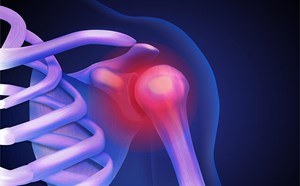
Hemarthrosis in Patients with Hemophilia
Justin Koss, DO
Inspira Medical Center Mullica Hill, Department of Emergency Medicine
C. Michael Lee, DO CAQSM
Kaiser Permanente Central Valley, Department of Emergency Medicine
Case:
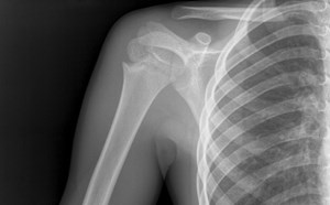
A 9-year-old male with a past medical history of hemophilia A presents with a right ankle injury after accidentally stepping into a divot while playing golf. Physical exam is notable for swelling around the ankle but no bony tenderness. X-ray reveals no acute bony abnormality but a joint effusion is noted.
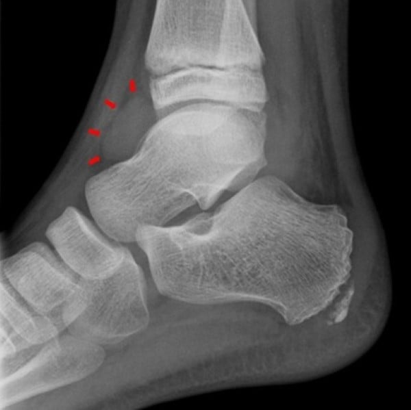 XR of the Right Ankle, Lateral View: Arrows point to the anterior portion of a joint effusion Case Courtesy of Dr. Andrew Dixon, Radiopaedia.org from case rID 10534. https://doi.org/10.53347/rID-10534
Background
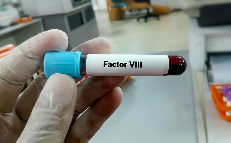
Hemophilia A and B are X-linked recessive bleeding disorders characterized by a defect or deficiency in factor VIII and IX, respectively. These disorders predispose patients to bleeding episodes, with hemarthrosis being a common occurrence. Hemarthrosis can occur spontaneously or due to trauma or injury. The most frequently affected joints are hinged joints (ankles, knees, and elbows).1
In patients with severe hemophilia, the first episode of hemarthrosis typically occurs between the ages of 1-5; thus, primary prophylaxis is normally initiated around this time in order to prevent or halt joint destruction.2 Without prophylactic treatment, patients with severe hemophilia may experience more than 30 episodes of hemarthrosis annually.2 Repeat episodes of hemarthrosis in these patients can lead to hemophilic arthropathy, a debilitating condition in which joints are impaired resulting in chronic pain and diminished quality of life.3 Additionally, these patients may also develop abnormalities in bone growth and limb length and are at increased risk of osteoporosis.2
Development of hemophilic arthropathy typically follows a progressive pathway where recurrent, untreated, or under-treated hemarthrosis leads to synovial inflammation, cartilage degeneration, and bone damage.3-5 Although the synovium of a joint has the ability to remove blood following an isolated episode of hemarthrosis, it typically requires time for the macrophages and other inflammatory cells to accomplish this task.5 Recurrent or persistent bleeding into the same joint, however, can result in a volume of blood that may overwhelm the body’s ability to clear it.5 Iron from the red blood cells collect as hemosiderin which triggers synovial inflammation.3,5 The inflamed synovium produces proteins such as cytokines that can then lead to cartilage and subchondral bone degradation.3-5
In the emergency department (ED), physicians are likely to encounter episodes of acute hemarthrosis which is characterized by pain and localized discomfort. If treatment is not initiated in a timely fashion, the joint may become significantly more painful or swollen. Because of the significant sequelae of hemarthrosis, it is prudent for an emergency medicine physician to familiarize themselves with diagnosing and treating patients with hemophilia who present with joint bleeding.
Diagnosis
Diagnosis of hemarthrosis is predominantly based on clinical presentation as there is currently no standardized protocol or validated diagnostic criteria.6 Although hemarthrosis can occur spontaneously in patients with hemophilia, most cases of hemarthrosis in patients with mild or moderate hemophilia are provoked by injury or trauma.7 Early symptoms of hemarthrosis may include joint fullness or stiffness, a tingling sensation, or pain while later symptoms may include swelling, warmth, decreased range of motion, tenseness, tenderness, inability to bear weight (for lower extremity joints), muscle spasm, a fixed flexed position, and a shine or erythema to the overlying skin.6
Imaging modalities such as x-rays (XR), computed tomography (CT), ultrasound (US), or magnetic resonance imaging (MRI) may be useful in visualizing a fluid collection. X-rays are not primarily used to diagnose or detect soft tissue injury or fluid collection although a joint effusion may be appreciated. A CT scan can provide more accurate details regarding the location and extent of effusion; however, it comes with an exposure to radiation and isn’t necessarily required for the sole purpose of identifying an effusion. Traditionally, MRI has been considered the “gold standard” when it comes to hemophilic arthropathy; however, MRI has significant disadvantages such as cost, accessibility or availability, and some patients may require sedation.8 One of the fastest and most reliable methods of detecting joint fluid is US, which is typically cost-effective, noninvasive, readily available, and does not convey radiation risk.9 Furthermore, US is extremely sensitive at detecting intra-articular blood at low concentrations and volumes and was able to discern between blood and non-blood fluid collections while MRI was not.10 However, US is operator dependent and would require additional training although it appears that with sufficient training even non-radiologists and non-physicians can perform US accurately.11
Although imaging studies can identify joint effusions, these modalities are not always definitive on the type of fluid that is present whether it is blood, synovial fluid, or pus. Arthrocentesis and subsequent fluid analysis would allow for better evaluation of the fluid. One study suggested that fluid aspiration can be useful for diagnostics when the origin of the effusion is unknown although aspiration would not be ideal when the fluid collection is secondary to trauma.12 However, in patients with hemophilia, routine arthrocentesis is not recommended.1,6,13,14
Treatment and Management
Because patients with hemophilia have a propensity for bleeding, special consideration must be taken in treating these patients when they present with concern for hemarthrosis. After ensuring stability of the patient (i.e. securing the airway if necessary, bleeding control), initiating replacement factor therapy and consultation with hematology.
Treatment should be based on clinical history and concern as physical exam may be unremarkable.15 Factor replacement should be administered as soon as possible upon suspicion or concern for a bleeding-related issue without waiting for confirmation of bleeding (e.g. labs or imaging).14-15 Patients may sometimes bring their own replacement factor with them to the hospital which can and should be administered if available. Ideally, the replacement factor should be given within 2 hours of bleeding onset.2 The goal is to increase the availability of the deficient factors as soon as possible in order to achieve hemostasis with a target level of 100% for major bleeds and 50% for minor bleeds.15 Although many different replacement factor concentrates exist, the dosing is the same: 50 U/kg of factor VIII or 100 U/kg of factor IX should be given to reach an availability level of 100%.15 When a patient’s baseline factor level is unknown, it should be assumed that the level is 0%, and if there is any doubt, full factor replacement should be administered.15 Depending on the response to the initial dose of treatment and with the assumption treatment consisted of standard half-life factor, patients with hemophilia A may require another dose 12 hours after the initial dose while patients with hemophilia B may require another dose 24 hours after initial dose.14 As repeat doses may be necessary after discharge from the ED, consultation with hematology to arrange for follow-up is important.
Aside from factor replacement, pain control, the RICE (Rest, Ice, Compression, and Elevation) approach, and rehabilitation are also important in helping patients recover.13-14 For analgesia, acetaminophen or opioids are reasonable options although long-term opioid use should be avoided.14 In general, aspirin and nonsteroidal anti-inflammatory drugs (NSAIDS) should be avoided although selective COX-2 inhibitors may be considered.2, 13,14 Although ice can decrease pain, there is evidence that lower intra-articular temperature may interfere with coagulation; thus, ice without direct skin contact should only be used for ~15 minutes at a time and only within the first 6 hours of injury.2,14
Protection or immobilization is also recommended and a “P” for protection is often added to the acronym RICE to create the acronym PRICE.14 Immobilization can be achieved with compression wraps or splinting although a circumferential wrap or splint should be avoided.2-13 If the bleed is in the hip, knee, or ankle joint, one day of bed rest is recommended, and the patient should be non-weight bearing for about 1 week with the resolution of pain and swelling guiding a return to weightbearing status.2,13,14 Elevation of the affected joint may help with swelling and can be utilized if tolerated by the patient.2,14 Although rest is important, prolonged immobilization can also negatively affect muscle strength so clinicians are encouraged to follow the POLICE (Protect, Optimal Loading, Ice, Compression, Elevation) approach in order to strike a balance between rest and early movement to avoid the consequences of immobilization while also minimizing rebleeding risks.2,14
The topic of arthrocentesis in these patients is controversial. One study found that joint aspiration led to faster resolution in bleeding, fewer days of treatment with factor replacement, faster pain relief, and earlier functional recovery and resumption of activities.16 At a 12 month follow-up, there was no difference between the control group and the group that underwent joint aspiration in terms of bleeding events, and there were no complications.16 Another study noted that patients undergoing joint aspiration had improved pain relief and functional improvement at 4 hours and 7 days.17 Ingram et al. found a statistically significant greater immediate range of motion in knees that underwent joint aspiration although by five days the range of motion between knees treated with and without aspiration were near equal.18
Although there may be some benefit to joint aspiration, the procedure carries risks including infection and bleeding. Per the World Federation of Hemophilia, arthrocentesis should not be routinely performed but can be considered if there are prolonged or worsening symptoms 24 hours after initial factor replacement treatment or if there is concern for infection.14 If arthrocentesis is performed, it should be performed when factor activity levels are adequate and under sterile conditions.14 For clinicians in the emergency department, it is probably best to avoid performing aspiration unless there is an emergent reason (such as concern for infection or compartment syndrome), and the procedure should only be performed under sterile conditions and after confirmation that factor replacement is adequate.14
Case Conclusion
The patient and his mother did not bring their supply of replacement factor as they came straight from the golf course. 50 U/kg of factor VIII replacement therapy was given to the patient in the ED in consultation with pediatric hematology. The patient was placed in a posterior short leg splint and discharged after pediatric hematology confirmed that they would call the patient and his mother the next morning to follow-up and provide guidance regarding a possible repeat dose of factor replacement therapy.
Summary
- Patients with hemophilia are at risk for bleeding with hemarthrosis being a common manifestation of the bleeding.
- Hemarthrosis can lead to hemophilic arthropathy which may result in debilitating pain and decreased function.
- Diagnosis is primarily clinical, and treatment with replacement factor should be given as soon as possible. Patients may require additional doses of the factor.
- Joint aspiration should not routinely be performed in the emergency department, but if joint aspiration is to be performed, it should be performed under sterile conditions and after confirmation of adequate factor levels/activity.
- To optimize patient care, it is important to find a balance between protection/rest and early mobilization/weightbearing.
References
- Srivastava A, Brewer AK, Mauser-Bunschoten EP, et Guidelines for the management of hemophilia. Haemophilia. 2013;19(1):e1-e47. doi:10.1111/j.1365-2516.2012.02909.x
- Lobet S, Hermans C, Lambert Optimal management of hemophilic arthropathy and hematomas. J Blood Med. 2014;5:207-218. Published 2014 Oct 17. doi:10.2147/JBM.S50644
- Melchiorre D, Manetti M, Matucci-Cerinic M. Pathophysiology of Hemophilic Arthropathy. J Clin Med. 2017;6(7):63. Published 2017 Jun doi:10.3390/jcm6070063
- Knobe K, Berntorp Haemophilia and joint disease: pathophysiology, evaluation, and management. J Comorb. 2011;1:51-59. Published 2011 Dec 27. https://doi:10.15256/joc.2011.1.2
- Pulles AE, Mastbergen SC, Schutgens RE, Lafeber FP, van Vulpen Pathophysiology of hemophilic arthropathy and potential targets for therapy. Pharmacol Res. 2017;115:192-199. doi:10.1016/j.phrs.2016.11.032
- Timmer MA, Pisters MF, de Kleijn P, de Bie RA, Fischer K, Schutgens RE. Differentiating between signs of intra-articular joint bleeding and chronic arthropathy in haemophilia: a narrative review of the literature. Haemophilia. 2015;21(3):289-296. doi:10.1111/hae.12667
- Zwagemaker AF, Kloosterman FR, Hemke R, et Joint status of patients with nonsevere hemophilia A. J Thromb Haemost. 2022;20(5):1126-1137. doi:10.1111/jth.15676
- Querol F, Rodriguez-Merchan The role of ultrasonography in the diagnosis of the musculo-skeletal problems of haemophilia. Haemophilia. 2012;18(3):e215-e226. doi:10.1111/j.1365-2516.2011.02680.x
- De la Corte-Rodriguez H, Rodriguez-Merchan EC, Jimenez-Yuste Point-of-care Ultrasonography in Orthopedic Management of Hemophilia: Multiple Uses of an Effective Tool. HSS J. 2018;14(3):307-313. doi:10.1007/s11420-018-9604-x
- Nguyen S, Lu X, Ma Y, Du J, Chang EY, von Drygalski Musculoskeletal ultrasound for intra-articular bleed detection: a highly sensitive imaging modality compared with conventional magnetic resonance J Thromb Haemost. 2018;16(3):490-499. doi:10.1111/jth.13930
- Acharya SS, Rule B, McMillan O, Humphries TJ. Point-of-care ultrasonography (POCUS) in hemophilia A: a commentary on current status and its potential role for improving prophylaxis management in severe hemophilia A. Ther Adv Hematol. 2017;8(4):153-156. doi:10.1177/2040620717690316
- Paschos NK, Giotis D, Abuhemoud K, Georgoulis AD. Effectiveness of aspiration in knee joint effusion management: a prospective randomized controlled Knee Surg Sports Traumatol Arthrosc. 2014;22(1):226-232. doi:10.1007/s00167-013-2379-1
- Rodriguez-Merchan, (2008) Articular bleeding (hemarthrosis) in hemophilia: An orthopedist’s point of view, 2nd Edition. Treatment of Hemophilia. Available at: https://www1.wfh.org/publication/files/pdf-1155.pdf.
- Srivastava A, Santagostino E, Dougall A, et WFH Guidelines for the Management of Hemophilia, 3rd edition. Haemophilia. 2020;26 Suppl 6:1-158. doi:10.1111/hae.14046
- Alblaihed L, Dubbs SB, Koyfman A, Long High risk and low prevalence diseases: Hemophilia emergencies. Am J Emerg Med. 2022;56:21-27. doi:10.1016/j.ajem.2022.02.045
- De la Corte-Rodriguez H, Rodriguez-Merchan EC, Alvarez-Roman MT, Martin-Salces M, Romero-Garrido JA, Jimenez-Yuste Accelerating recovery from acute hemarthrosis in patients with hemophilia: the role of joint aspiration. Blood Coagul Fibrinolysis. 2019;30(3):111-119. doi:10.1097/MBC.0000000000000803
- Rai AK, Mohanty SS, Rathod TN, Kamble P, Keny SA, Kothari Outcome of Joint Aspiration in Acute Knee Haemarthrosis in a Haemophilic Joint: A Prospective Randomised Controlled Trial in 120 Patients in a Tertiary Haemophilia Care Centre. Indian J Orthop. 2022;56(12):2060-2065. Published 2022 Sep 16.
a. doi:10.1007/s43465-022-00745-x - Ingram GI, Mathews JA, Bennett A controlled trial of joint aspiration in acute haemophilic haemarthrosis. Br J Haematol. 1972;23(6):649-654. doi:10.1111/j.1365-2141.1972.tb03480.x