1st Place Emage Winner: A Morel-Lava WHAT!?!
Nithin Ravi, MD
Tien Vu, MD
Samuel Lam, MD, FACEP
Children's Hospital of Colorado, Aurora, CO
Case
A 16-year-old otherwise healthy male presents with right lower extremity swelling with overlying skin changes 12 days status-post motor vehicle collision (MVC); the patient was unrestrained in a rear passenger seat when involved in the single-car rollover MVC traveling at 80 mph. He was ejected from the vehicle and his right leg was pinned under the vehicle at the site of current concern.
He was seen at an outside emergency department (ED) where he received a full trauma evaluation, including x-rays of femur, tibia, and fibula which were all reportedly negative. He was discharged from the ED ambulatory. Four days later, he noticed darkening of the skin that he thought was a scab but progressed throughout the week with worsening ecchymoses and swelling. He has had no difficulty with ambulation, no pain with range of motion of the leg, only mild tenderness to palpation, mild numbness on the superficial skin just surrounding the area, and scant blood draining from the dark lesion intermittently, with no purulent drainage reported, and no fevers, or other systemic symptoms.
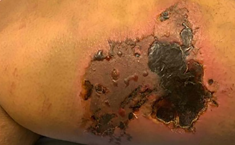
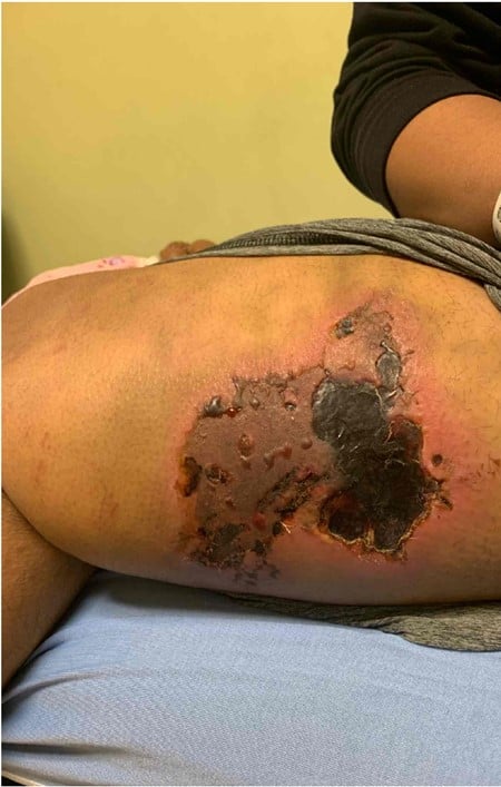
Physical examination showed a large (~ 7 cm x 5 cm) area of dark skin on the medial thigh with a coarse, leathery, texture (Figure 1). Upon palpation, small speckles of blood seeped through the lesion. It has surrounding erythema, but no warmth, and minimally tender to palpation. Ecchymosis and swelling extend from the inguinal canal down to the knee on the medial aspect of the thigh. The surrounding skin is taut, but compressible.
Workup
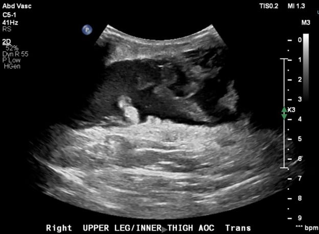
Ultrasound showed a large, elongated fluid collection within the subcutaneous soft tissues of the upper medial thigh at the site of concern measuring 24 cm longitudinal x 2.8 cm transverse, characterized by irregular margins, low level internal echoes that swirled upon compression, and irregular linear internal echogenic foci that may have reflected fat. These findings were consistent with a Morel-Lavallée degloving injury.
Management
Pediatric surgery was consulted to report the Morel-Lavalleé finding on ultrasound, to which the surgery resident responded, "A Morel-Lava WHAT?!?!" The patient was taken to the operating room (OR) for incision and drainage, washout, debridement of necrotic tissue, and wound vacuum-assisted closure (VAC) placement. A total of 1200 mL old blood and clot was drained from the anteromedial thigh. During the 1-week admission, he underwent two trips back to the OR due to re-accumulation of fluid in the underlying space, requiring wound VAC replacement and further debridement. White blood cell count was 13.71 and wound culture grew methicillin-sensitive Staphylococcus aureus (MSSA). The patient was treated with cefazolin and transitioned to cephalexin at the time of discharge, with a wound VAC in place.
Clinical Pearls
A Morel-Lavallée lesion is a post-traumatic, internal closed degloving injury. The mechanism of injury is typically a high velocity trauma with shearing forces or a crush injury. During the injury, the deep fascial layers become separated from the skin and superficial fascia. This creates an open space in the pre-fascial plane for blood and fluid to collect from injured capillary vessels and/or lymphatics during the trauma. The hemosiderin from the blood creates an inflammatory process which over time leads to an outer fibrous capsule formation with the internal space filled with necrotic tissue, blood products, fibrin, and debris. This fluid stasis can be a nidus for infection and must be considered in patients with fevers or signs and symptoms of a skin and soft tissue infection.
These injuries are typically associated with fractures of the proximal femur, pelvis, or acetabulum. They are most frequently present between the knee and hip, since the overlying skin is mobile and the underlying fascia is tough making it easier for these planes to separate. These injuries are often missed on initial presentation due to the lack of external skin findings, but progress over weeks to months. The diagnosis is typically made clinically on re-presentation. Skin changes can vary and include ecchymosis, swelling, cracking, drying, abrasions, or frank necrosis from underlying pressure. Cutaneous anesthesia is common due to damage of the subdermal afferent nerves. A diagnosis can be made clinically, but is often confirmed on ultrasound, computed tomography (CT), or magnetic resonance imaging (MRI).
There are no gold standard guidelines for management of Morel-Lavallée lesions; however, four approaches exist:
- Conservative management: Application of compression bandages. Only indicated if the lesion is small and there is no capsule formation.
- Percutaneous aspiration: Recurrence rate is high, particularly with lesions containing more than 50 mL of volume.
- Sclerodesis: Mechanism of action is cellular destruction on the periphery of the lesion, resulting in fibrosis. Efficacy of this method has been reported at 95.7%.
- Open drainage and mass resection.
References
- Agrawal U, Tiwari V. Morel Lavallee Lesion. [Updated 2023 Jan 21]. In: StatPearls [Internet]. Treasure Island (FL): StatPearls Publishing; 2023 Jan. Available from: https://www.ncbi.nlm.nih.gov/books/NBK574532/