2021 1st Place Emage Winner: Abdominal Distention: Prepare for the Worst
Case Presentation
CC: Abdominal pain
History and Examination:
A 19-year-old female with past medical history of aerophagia, autism, developmental delay, seizure disorder, chronic constipation presented to the ED complaining of abdominal pain. Parents reported 2-3 days of worsening abdominal distention and pain, decreased stooling, and decreased activity. Parents stated that this is the most distended she has ever been. She had prior hospitalizations for aerophagia and distention, and usually decompressed spontaneously or with rectal tube. Her ROS was positive for abdominal pain and nausea, and negative for fever, vomiting, diarrhea, hard stool or blood in the stool. Previous surgeries included myringotomy with bilateral tubes. She lives at home with her parents and is up-to-date on vaccinations. She has multiple food allergies. They are compliant with her home medications.
On exam, she appeared ill and acutely distressed. Her vitals were HR 136, BP 139/108, T 37C, RR 16, and Spo2 on room air of 100%. She had dry mucous membranes, and her abdomen was severely distended, tense, tender, and tympanic with hypoactive bowel sounds. Her capillary refill was delayed to 3-4 seconds.
Immediate ED Interventions:
- Inserted IV, labs sent, gave IV fluid bolus 20/cc/kg of NS and IV Zofran
- Obtained X-rays: cross table lateral, abdominal series, chest
- Inserted NGT on continuous suction – had minimal output and no reduction in abdominal distention
- Placed oral / nasal ETCO2
- After initial interventions, prepared for rectal tube placement with ketamine
ED course:
- Placed red rubber catheter and 48F chest tube inserted rectally
- Unsuccessful in decompression, planned for OR
Then became agitated and obtunded
developed desaturations, worsening tachypnea and tachycardia, agonal respirations, and weak radial and femoral pulses - Started BVM for respiratory failure
- HR dropped to 50bpm with weak radial pulse and pulseless femoral —> CPR initiated
- Performed successful endotracheal intubation then noted to have normal palpable radial pulses, with persistently absent femoral pulses
- Tibial IO was placed for difficult IV access
- Despite securing airway, pt. noted to have persistent hypoxia with spo2 in 70%s
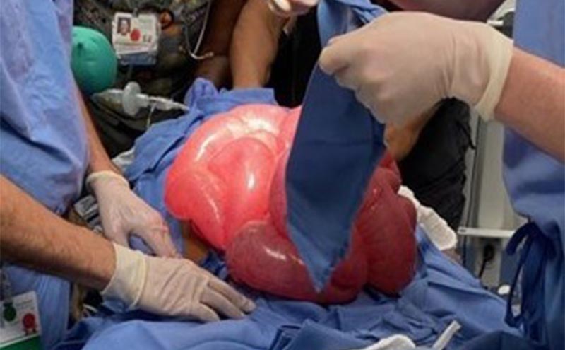
- Performed decompressive laparotomy in the ED trauma bay, see Figure 4 Spo2 then normalized and pt. regained femoral pulses
transported to the OR covered in moist sterile towels
Hospital course:
The patient was transported to the OR for an emergent exploratory laparotomy. She was noted to have a volvulus that involved the cecum and adjacent bowel. General surgery performed an enterotomy in both the small and large bowel, and decompressed and re-oriented the bowel. She was admitted to the PICU, and eventually was transferred to the floor. Her course was complicated by an additional exploratory laparotomy for increasing abdominal distention and free air. She was eventually discharged home.
Discussion:
Due to severe abdominal distention from aerophagia, the patient developed acute abdominal compartment syndrome (ACS) in the ED, and subsequent respiratory and cardiac arrest necessitating intubation, CPR, bedside decompressive laparotomy, further surgical exploration in the OR, and PICU admission. The etiology of her ACS was increasing abdominal distention and worsening intra-abdominal hypertension (IAH) from intra-luminal air secondary to her aerophagia and a concomitant cecal volvulus.
ACS results from increasing or persistent IAH, and can present with a broad spectrum of signs and symptoms. These could include abdominal pain and distention, nausea and vomiting, altered mental status, hemodynamic instability, and even shock.
In the ED setting, it can be difficult to recognize ACS as the primary cause of multi-organ dysfunction. ACS is diagnosed most accurately by measuring intra-abdominal pressures, but in the ED setting, the physician must have high clinical suspicion as measuring intra-abdominal pressure is not always feasible. ED management is directed towards supportive care, decompressing the abdomen to decrease IAH in effort to prevent the development of ACS, and arranging surgical evaluation and intervention as needed.
The etiologies of pediatric ACS are thought to be similar to those in adults. These include bowel obstruction, traumatic intra-abdominal injuries and intra-abdominal bleeding, recent liver transplant, aerophagia, abdominal mass, and increased abdominal perfusion or re-perfusion often associated with fluid resuscitation or in the post-surgical setting.1 In the pediatric population it is usually associated with intra-abdominal trauma, surgical abdominal emergencies, post-surgery (intra-abdominal or cardiac), fluid resuscitation in burns or sepsis, and post CPR. If untreated in the pediatric population, it causes multiple organ failure, and has a 90%-100% mortality rate.3 ACS is usually refractory to medical management.2 Even with surgical decompression, the literature describes a persistently high mortality rate, ranging from approximately 40-60%.4,5
As intra-abdominal pressure rises, progressive ischemia to the intra-abdominal organs can lead to acute organ dysfunction, including acute kidney injury, ischemic bowel and perforation, and ischemic hepatitis.1,6 With ACS, there is decreased venous return to the heart and therefore decreased cardiac output, leading to shock and ultimately asystole. With increasing abdominal distention, there is decreasing lung capacity and worsening ventilation, potentially precipitating respiratory and cardiac arrest. ACS can lead to multi-compartment syndrome, such as thoraco-abdominal compartment syndrome, and poly-compartment syndrome (neuro-thoraco-abdominal compartment syndrome). Understanding the pathophysiology of IAH causing ACS is crucial to stabilizing the patient while arranging emergent surgical intervention.
Conclusion:
Acute abdominal compartment syndrome in the pediatric population has fewer case reports and studies in the literature in comparison to the adult population. We hope our case report sheds light on this rare and potentially fatal syndrome. Further research regarding early intervention of IAH in the pediatric population and ED setting could help prevent the development of ACS and shock.
Take home points:
- Early signs and symptoms of IAH and ACS include abdominal pain, nausea, vomiting, abdominal distention, and can progress to difficulty breathing, altered mental status, cardiovascular compromise, and shock.
- The pathophysiology of ACS leads to multiple organ system dysfunction and failure and is eventually fatal if untreated.
- In the ED, patients with suspected IAH should be decompressed if appropriate.
- Patients with ACS in the ED should be resuscitated appropriately while arranging emergent surgical management.
Statement and references:
The authors declare that they have no competing interests.
- Newman RK, Dayal N, Dominique E. Abdominal Compartment Syndrome. [Updated 2020 Jul 10]. In: StatPearls [Internet]. Treasure Island (FL): StatPearls Publishing; 2021 Jan-. Available from: https://www.ncbi.nlm.nih.gov/books/NBK430932/
- Cheatham ML. Abdominal compartment syndrome: pathophysiology and definitions. Scand J Trauma Resusc Emerg Med. 2009;17:10. Published 2009 Mar 2. doi:10.1186/1757-7241-17-10
- Newcombe J, Mathur M, Ejike JC. Abdominal compartment syndrome in children. Crit Care Nurse. 2012 Dec;32(6):51-61. doi: 10.4037/ccn2012761. Phttps://link.springer.com/content/pdf/10.1007/s00134-013-2906-z.pdfMID: 23203955.
- Ejike JC, Newcombe J, Baerg J et al (2010) Understanding of abdominal compartment syndrome among pediatric healthcare providers. Crit Care Res Pract.
- Je B, Kim H, Horn P. Abdominal Compartment Syndrome in Children: Clinical and Imaging Features. American Journal of Roentgenology. 2019;212: 655-664. 10.2214/AJR.18.20119
- Gurel A. Acute kidney injury due to abdominal compartment syndrome caused by duodenal metastases of prostate cancer. Clin Case Rep. 2015;3(7):629-631. doi:10.1002/ccr3.305
- Beck, Raphael MD; Halberthal, Michael MD; Zonis, Ze’ev MD; Shoshani, Gideon MD; Hayari, Lili MD; Bar-Joseph, Gad MD Abdominal compartment syndrome in children,
- Pediatric Critical Care Medicine: January 2001 - Volume 2 - Issue 1 - p 51-56
- Kirkpatrick, A.W., Roberts, D.J., De Waele, J. et al. Intra-abdominal hypertension and the abdominal compartment syndrome: updated consensus definitions and clinical practice guidelines from the World Society of the Abdominal Compartment Syndrome. Intensive Care Med 39, 1190–1206 (2013).
- Chiles KT, Feeney CM. Abdominal compartment syndrome successfully treated with neuromuscular blockade. Indian J Anaesth. 2011;55(4):384-387. doi:10.4103/0019-5049.84867
ED abdominal images and workup:
Significant lab results included a non-anion gap metabolic acidosis.
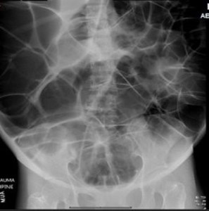
Figure 1. Anteroposterior (AP) abdominal plain film obtained in the ED.
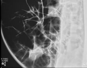
Figure 2. supine cross table abdominal plain film obtained in the ED.
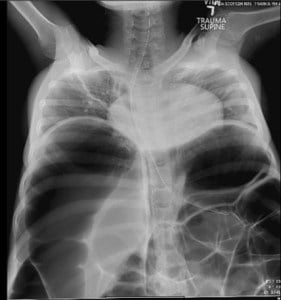
Figure 3. AP abdominal plain film obtained in the ED.
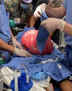
Figure 4. Decompressive laparotomy in the ED trauma bay
Dr. Caitlin Mueller, DO, PGY3 EM
Dr. Mona Kulkarni, MD, PEM
Dr. Thomas Abramo, MD, PEM