Critical Care Updates: Two Views to Save Lives: Transesophageal Echocardiography for Peri-Arrest and Cardiac Arrest Patients
Although transesophageal echocardiography (TEE) has long been used to help diagnose valvular heart disease, wall-motion abnormalities, strain, and cardiac outflow, it has been recently shown to have significant benefits in the management of emergency department (ED) patients in cardiac arrest 1,2 Here is a useful video primer on the value of TEE in cardiac arrest patients: Watch Video.
Transthoracic echocardiography during cardiac arrest can be technically difficult and often leads to long interruptions in chest compressions.3 By providing an unobstructed view of the heart and the great vessels, TEE allows clinicians to accurately evaluate cardiac function and the quality of chest compressions as well as perform resuscitative measures with the probe still in place. TEE can also be useful in the unstable, intubated ED patient in order to obtain more information about their cardiac function, fluid status, the etiology of their shock state, and previously unknown pathologies (e.g. aortic dissection or pulmonary embolism).4,5
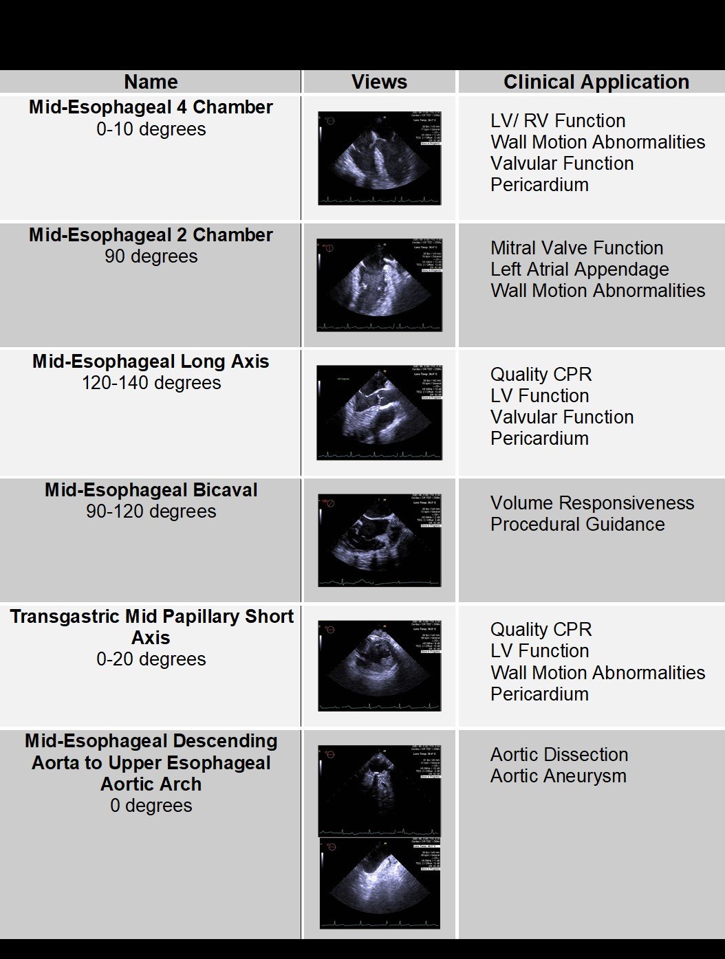
The 2017 ACEP guidelines recommend a four-view limited point of care exam for the patient that presents to the ED in cardiac arrest.6
These four views are the:
- Midesophageal 4-Chamber view at 0 degrees
- Insert the probe at 0 degrees while watching the screen until all 4 chambers are seen. Anteflexion of the probe tip may be required to get the probe into the esophagus.
- Midesophageal Long-Axis view at 120 degrees
- From View 1 increase the omniplane from 0 Degrees to 120 degrees while keeping the LV in the center of the screen.
- Midesophageal Bicaval view at 120 degrees
- Rotate the probe to the patient’s right side from the Midesophageal Long Axis view
- Transgastric Mid-Papillary Short-Axis view at 0 degrees
- From View 3 return the omniplane back to 0 degrees and rotate back to the patient’s left to obtain the midesophageal 4 chamber view. Gently advance the probe into the stomach. Once in the stomach use the large wheel to ante-flex the probe tip.
Quick Six
In addition to the published views for TEE in cardiac arrest, we propose two supplemental views that provide additional information in both the peri-arrest and cardiac arrest patient and can be used in settings beyond the ED.7 These include the Mid Esophageal 2-Chamber View at 90 degrees and the Descending Aorta and Aortic Arch View at 0 degrees.
How To Obtain The Supplemental Views
These two additional views are easily obtained. The first view is the Midesophageal 2 chamber view which is obtained by simply increasing the omniplane to 90 degrees from the midesophageal 4 chamber view at 0 degrees. This will afford the examiner views of the anterior and inferior walls of the left ventricle as well as the mitral valve. This is clinically useful in the setting of possible acute myocardial infarction and also provides a great view of the mitral valve allowing one to assess for valvular dysfunction or vegetation. The second view can be obtained from the Transgastric Mid Papillary Short Axis view by returning the omniplane to 0 degrees and rotating the probe to patient’s left. Slowly withdraw the probe until the descending aorta is seen. One then continues withdrawing the probe while keeping the aorta in view, until the aortic arch is reached in the upper esophagus. To get a more complete view of the aortic arch one must rotate the probe slightly towards the patient’s right. This extra view is simple to acquire and adds a great deal of information about the aorta, assessing for dissection or aneurysm, while one is removing the probe.
References
- Arntfield RT, Millington SJ. Point of care cardiac ultrasound applications in the emergency department and intensive care unit--a review. Curr Cardiol Rev. 2012 May; 8(2):98-108.
- CPR reexamined: What we know, what we don't know and a tool that can help. https://www.resuscitativetee.com
- van der Wouw PA, Koster RW, Delemarre BJ, et al. Diagnostic accuracy of transesophageal echocardiography during cardiopulmonary resuscitation. J Am Coll Cardiol. 1997 Sep;30(3):780-783.
- Huis In‘t Veld MA, Allison MG, Bostick DS, et al. Ultrasound use during cardiopulmonary resuscitation is associated with delays in chest compressions. Resuscitation. 2017 Oct;119:95-98
- Jones AE, Tayal VS, Sullivan DM, et al. Randomized, controlled trial of immediate versus delayed goal-directed ultrasound to identify the cause of non-traumatic hypotension in emergency department patients. Crit Care Med. 2004 Aug;32(8):1703-1708.
- Jones AE, Craddock PA, Tayal VS, et al. Diagnostic accuracy of left ventricular function for identifying sepsis among emergency department patients with non-traumatic symptomatic undifferentiated hypotension. Shock. 2005 Dec;24(6):513-517.
- American College of Emergency Physicians. Guidelines for the use of transesophageal echocardiography (TEE) in the ED for cardiac arrest. [policy statement]. Ann Emerg Med. 2017; 442-445
- Arntfield R, Pace J, McLeod S, et al. Focused transesophageal echocardiography for emergency physicians-description and results from simulation training of a structured four-view examination. Crit Ultrasound J. 2015 Dec; 7(1):27.
Maria O’Rourke, MD
Byron Mendenhall, MD
Elisa M. Aponte, MD
Kaweah Delta Medical Center