Diagnosis of Tibial Tubercle Fracture on Point-of-Care-Ultrasound
Emily Cen, MD, Maimonides Medical Center
Irina Sanjeevan, MD, Maimonides Medical Center
Leily Naraghi, MD, Maimonides Medical Center
Introduction
A tibial tubercle fracture is an uncommon pediatric fracture accounting for < 1% of epiphyseal fractures and 3% of proximal tubercle avulsion fractures.1 It presents as knee pain and occurs more commonly in males, adolescents, and those who participate in athletic activities such as basketball or sprinting. It is important to diagnose this injury to avoid complications such as compartment syndrome, atrophy of the quadriceps, or loss of knee flexion.2 Existing literature on the utility of ultrasound in the diagnosis of this injury is limited to one case report and a single-center study, but does support its use.3,4 This case report aims to highlight the utility of point-of-care ultrasound (POCUS) in the rapid diagnosis of traumatic knee pathologies including tibial tubercle fractures in the emergency department (ED).
Case Presentation
A 16-year-old male with no significant past medical history presented to the ED with knee pain that began suddenly while playing basketball. He had led his jump with his right leg, and then reportedly "felt a pop" in his left knee while midair. He was unable to ambulate after the injury. He denied any other complaints including head strike or loss of consciousness.
His initial vital signs were as follows: temperature 37.1 C, blood pressure of 116/85 mmHg, heart rate of 88 beats/minute, respiratory rate of 18 breaths/minute, and oxygen saturation of 99% on room air. On physical exam, he was found to have significant swelling and diffuse tenderness to his left knee extending to his mid-shin, with decreased range of motion of the knee. He was unable to fully extend the knee either actively or passively. The remainder of his physical exam including the neurovascular exam of the affected extremity was otherwise unremarkable.
POCUS of the left knee was performed, which was concerning for a patellar tendon rupture and a joint effusion. (Figure 1 and Video 1) In addition, there was posterior acoustic shadowing obscuring part of the patellar tendon concerning for an avulsed bony fragment. (Figure 2) X-rays were performed, which confirmed the suspected diagnosis of a left tibial tubercle avulsion. (Figure 3) The patient was ultimately taken to the operating room for an Open Reduction and Internal Fixation (ORIF) by Orthopedic Surgery and discharged home the following day in a knee brace.
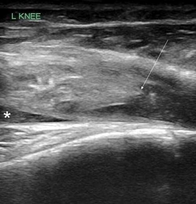
Figure 1. Ruptured patellar tendon (arrow) with joint effusion (*) seen in longitudinal view
Video 1. Evaluation of the subpatellar space in transverse plane using compression that demonstrates swirl sign, suspicious for hemarthrosis or lipohemarthrosis.
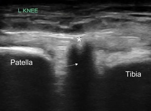
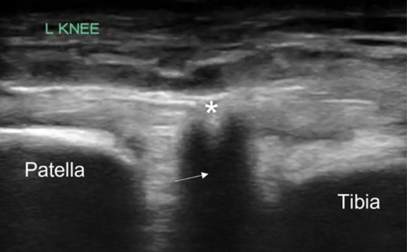
Figure 2. Patellar tendon with focus of acoustic shadowing (arrow) concerning for avulsed bony fragment (*)
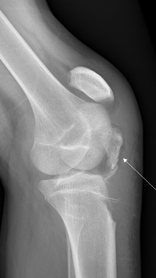
Figure 3. Lateral knee x-ray showing tibial tubercle avulsion (arrow)
Discussion
Tibial tubercle fractures typically occur in adolescent males after jumping or sprinting. This pathology thought to be due to relative skeletal immaturity relative to anterior cruciate ligament (ACL) strength during strenuous activity.5,6 Proper diagnosis of the fracture is crucial in avoiding complications such as compartment syndrome, atrophy of the quadriceps, or loss of knee flexion due to associated ligament injury.6 These fractures are typically diagnosed on X-ray, but can be missed in skeletally immature adolescents or if the avulsed fragment is small.3,4 Second-line imaging such as computed tomography (CT) or magnetic resonance imaging (MRI) may be needed to confirm the suspected diagnosis. However, MRIs may be very difficult to obtain emergently (or at all) in the ED, and CTs expose the patients to ionization radiation. We propose that POCYS should be considered to expedite detection of these fractures when evaluating patients with acute knee injuries.
In our case report, we successfully identified key features suggestive of tibial tubercle fractures: disruption of the patellar tendon, posterior acoustic shadowing of a bone fragment, and a large joint effusion. To perform knee ultrasound, the linear probe should be used and placed inferior to the patella in sagittal plane. The patellar tendon should be visualized in two planes, fanning through it completely (Video 1), and compared to the contralateral normal knee. (Figure 4) Abnormal findings suggestive of epiphyseal fractures include disruption of the tendon, an increased hypoechoic space adjacent to or posterior to the tendon (suggestive of either hemarthrosis or a hematoma), or a hypoechoic zone (ie, posterior acoustic shadowing due to an avulsed bony fragment).3,4
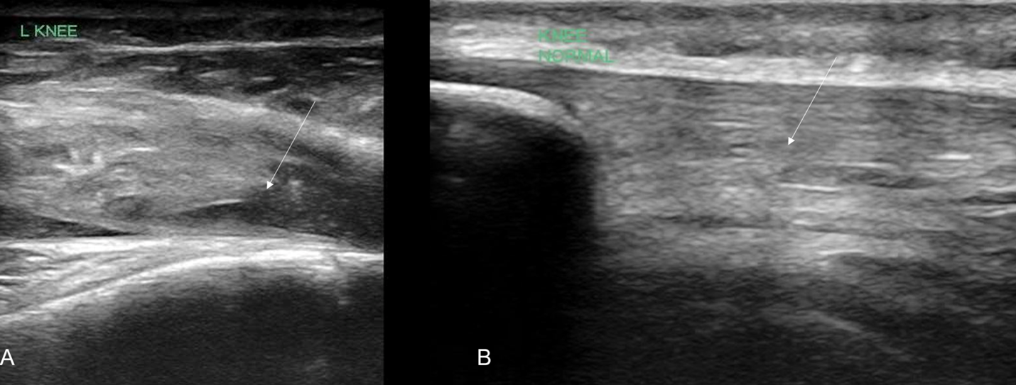
Figure 4: A) Ruptured tendon (arrow) in comparison against (B) contralateral knee indicating the normal continuous tendon fibers (arrow)
Conclusions
Our case report demonstrates the utility of POCUS in the evaluation of acute knee injury in adolescents. Musculoskeletal POCUS can be used to further expedite and facilitate proper diagnosis of tibial avulsion fractures in the ED, thus avoiding its possible complications and providing these patients with an appropriate plan of care. To our knowledge, this POCUS modality is currently underutilized, and this case highlights the need for further studies looking at musculoskeletal POCUS for timely diagnosis of knee injuries in the ED.
References
- Bolesta MJ, Fitch RD. Tibial tubercle avulsions. J Pediatr Orthop. 1986;6(2):186-92. doi:10.1097/01241398-198603000-00013
- Cole WW 3rd, Brown SM, Vopat B, et al. Epidemiology, diagnosis, and management of tibial tubercle avulsion fractures in adolescents. JBJS Rev. 2020;8(4):e0186. doi:10.2106/JBJS.RVW.19.00186
- Acuna J, Situ-LaCasse E, Jamplis RP, Amini R, Adhikari S. Point-of-care ultrasound evaluation of tibial avulsion fractures. Cureus. 2018 May 23;10(5):e2677. doi: 10.7759/cureus.2677. PMID: 30050732; PMCID: PMC6059524.
- Lazović D, Wegner U, Peters G, et al. Ultrasound for diagnosis of apophyseal injuries. Knee Surg Sports Traumatol Arthrosc. 1996;3:234-7. doi:10.1007/BF01466625
- Frey S, Hosalkar H, Cameron DB, et al. Tibial tuberosity fractures in adolescents. J Childr Orthop. 2008;2:469-74. doi:10.1007/s11832-008-0131-Z
- Pretell-Mazzini J, Kelly DM, Sawyer JR, et al. Outcomes and complications of tibial tubercle fractures in pediatric patients: a systematic review of the literature. J Pediatr Orthop. 2016;3:440-6. doi:10.1097/BPO.0000000000000488 https://pubmed.ncbi.nlm.nih.gov/25887827/