 American College of Emergency Physicians

Transforming Ultrasound Simulation into ABEM-Level Reasoning
Chintan Harish Kumar, MD¹
Vanessa Guillen, MD¹
Solomon Sebt, MD¹
Francisco Javier Andrade Jr., MD, FPD-AEMUS2
Trent She, MD, FACEP, FPD-AEMUS3
Drew Frey, MD, FPD-AEMUS4
Edmund Hsu, MD, MSIDT, FPD-AEMUS5
¹Department of Emergency Medicine, Eisenhower Health, Rancho Mirage, California
2Department of Emergency Medicine, Abrazo Health, Phoenix, Arizona 85013
3Department of Emergency Medicine, Hartford Hospital, Hartford, CT 06106
4Department of Emergency Medicine, Southern Illinois University, Springfield Il 62794
5Department of Emergency Medicine, University of California Irvine, Orange, CA 92868
This article was written in collaboration between the ACEP Emergency Ultrasound Section - Ultrasound Simulation and Fellow Education Subcommittees. Thank you to all involved and we hope this will provide help in preparing your residents for the upcoming ABEM Competency Exam.
A Quick Thought Before We Start
In many introductory POCUS curricula, early training appropriately emphasizes image acquisition technique and recognition of normal sonographic anatomy before advancing to diagnostic integration.1,2 But in simulation, the most valuable moments occur when the learner must interpret, act, and communicate, all in real time. Those moments shift ultrasound from a purely technical skill to a reasoning exercise. Recognizing this, we began exploring how simulation can also serve as a deliberate training ground for structured clinical reasoning, the same kind tested on the ABEM oral exam.
Why This Approach Works
Ultrasound simulation already demands many of the cognitive tasks required on the new ABEM exam: acquire quality views, describe relevant anatomy and interpret pathologic images.3,4 By reframing existing ultrasound SIM cases into ABEM-style oral scenarios, educators can train both diagnostic precision and structured verbal reasoning simultaneously—without needing new cases or added curricular time.5,6
Connecting Simulation with Exam Mindset
Ultrasound simulation inherently trains the four ABEM domains - Data Gathering, Synthesis, Management, and Communication, but often without explicitly naming them. By converting a SIM into an ABEM-style oral case, we make those domains visible and deliberate. Trainees practice not only what to see, but how to think and lead when the stakes are high.
A Brief Word on the ABEM Certifying Exam
ABEM’s new certifying exam replaces the long-standing oral exam beginning in 2026.4 After passing the Qualifying Exam, candidates will complete an in-person certifying exam held in Raleigh, North Carolina.6 The facility is the same high-volume simulation center used by multiple specialty boards—including Anesthesiology, Oral & Maxillofacial Surgery, a`nd Urology—and is specifically designed to accommodate 3,000+ emergency medicine candidates per year.7
The move to in-person testing reflects ABEM’s goal to more accurately replicate real emergency department practice, integrating realistic settings, standardized patients, ultrasound machines, and hands-on procedural stations.3,5 ABEM consulted over 4,000 stakeholders (residents, program directors, EM organizations, the public) and conducted multiple beta tests to refine exam structure, fairness, and clarity.4
The exam consists of two major components:6
1. Clinical Decision-Making Cases
- Structured interview cases assessing diagnostics, differential diagnosis, and management plans.4
- Prioritization cases mirroring real triage decisions and managing multiple sick patients simultaneously.5
2. Communication & Procedural Cases
- Delivering bad news
- Managing conflict with consultants or patients
- Patient-centered shared decision-making
- Reassessment when a patient clinically deteriorates.4
- Performing or demonstrating critical EM procedures using low-fidelity task trainers4,6
- Specifically for ultrasound: requiring a choice in probe selection, directing adjustments to depth/gain/preset settings, acquiring images on standardized patients, and interpreting pathology.5,6,8
- Interestingly, Emergency Medicine is among the first specialties to incorporate direct assessment of procedural performance into board certification in this format.6
The exam is criterion-referenced, meaning passing standards are based on what practicing EM physicians consider competent—not on a curve or preset failure rate.6 A notable addition is a dedicated point-of-care ultrasound (POCUS) case to test applicants. Candidates are not expected to be familiar with the specific machine’s controls but candidates must be able to verbalize anatomy during image acquisition,5 select appropriate probes, direct adjustments in depth or gain,5,6 interpret ultrasound clips5,6 and interact professionally with standardized patients.7The case is intended to mimic the use of POCUS in the clinical care of a patient who presents to the emergency department.
Additionally, the new ABEM Certifying Exam is designed not to test recall but to evaluate how physicians think, synthesize information, manage uncertainty, and communicate decisions in real time.3 The exam’s ultrasound component evaluates skills across applications listed in the EM Model of Clinical Practice Section 19.5.8 Each case probes four cognitive domains: Data Gathering, Synthesis, Management, and Communication - that mirror the decision-making rhythm of actual emergency care. Success depends on clarity, prioritization, and composure rather than memorized answers. It’s a test of reasoning under pressure, which is the same high-cognitive environment where ultrasound simulation excels. Both demand rapid interpretation, decisive action, and effective leadership in evolving scenarios.
Why It’s Worth Doing
Efficiency
Uses existing ultrasound SIMs to build ABEM-aligned reasoning skills without creating new cases.3
Relevance
Directly supports skills tested on the certifying exam, including ultrasound acquisition, interpretation, procedural readiness, triage decisions, and communication.3,4,8
Faculty Growth
Develops examiner-style questioning, structured prompting, and competency-based feedback—skills aligned with ABEM’s assessment philosophy.4,6
Learner Growth
Builds confidence, structure, and adaptability—skills essential for ultrasound OSCEs, procedural evaluations, and communication stations.4,5,7
Fairness & Accessibility
ABEM incorporates psychometric validation, inter-rater reliability checks, and accommodations for candidates with disabilities.4
- Efficiency: Leverages existing simulation content without increasing preparation time.
- Relevance: Aligns ultrasound education with ABEM’s structured reasoning framework.
- Faculty Growth: Strengthens examiner-style questioning and structured feedback.
- Learner Benefit: Builds confidence, clarity, and adaptability under pressure.
How to Translate a SIM Case into an ABEM Case
|
SIM Element |
ABEM Equivalent |
How to Adapt |
|
Scenario Setup |
Opening Stem |
Condense into a one-line clinical snapshot3,4 |
|
Learning Points |
Decision Points |
Identify 3–4 critical reasoning junctures4 |
|
Ultrasound Images |
Prompted Findings |
Ask: “You obtain this view. What do you see?”5,6 |
|
Debrief Questions |
Examiner Prompts |
Turn reflective questions into real-time follow-ups3 |
|
Checklist |
Critical Actions |
Map to ABEM’s domains + ultrasound OSCE expectations5,7 |
ABEM ultrasound cases require 2–3 image acquisitions and 2–3 clip interpretations within 10 minutes, with examiners allowed to interrupt to maintain timing.7
SIM Case Elements vs. ABEM Exam Equivalents
Case Example 1: EFAST in the Actively Unstable Trauma
Scenario:
A 27-year-old male is brought in after a high-speed rollover MVC. He is pale, diaphoretic, and hypotensive (BP 84/50, HR 132). There’s obvious abdominal distention and a seat-belt sign across the lower abdomen. Two peripheral IV attempts have failed, and the blood bank reports that crossmatched units are delayed.
Goal:
Integrate ultrasound findings with trauma resuscitation priorities in a rapidly decompensating patient.
Ultrasound Findings:
Initial RUQ EFAST window shows a faint hypoechoic stripe near Morrison’s pouch (equivocal for free fluid).
 Positive FAST - RUQ. Used with permission of the POCUS Atlas. Content License. Image source: https://www.thepocusatlas.com/trauma/bj4stp5hebk8ifh6pvwtirmswszk2q
For additional visual examples of free fluid patterns in Morrison’s pouch, see The POCUS Atlas Trauma Collection: https://www.thepocusatlas.com/bb-trauma
Examiner Prompts (ABEM Domains and Rationale)
- Airway and Initial Actions: “You’re leading the trauma team. What are your first three steps?”
Ideal Response: Ensure airway control, initiate massive transfusion protocol, and obtain vascular access (introducer or IO) while assigning roles. (Management + Communication) - Equivocal FAST: “Your RUQ view is indeterminate due to bowel gas. What’s next?”
Ideal Response: Repeat the RUQ or shift to a left-sided or suprapubic view; correlate with clinical picture rather than relying solely on a single window. (Data Gathering + Synthesis) - Competing Injuries: “Pelvic X-ray shows an open-book fracture. How does that alter priorities?”
Ideal Response: Apply pelvic binder, continue MTP, reassess for dual bleeding sources; balance time to OR vs. IR depending on local resources. (Synthesis + Management) - Limited Resources: “Surgery is occupied and blood products are still in transit. The patient’s BP is 80/50 after one unit of uncrossmatched O-negative. What’s your next move?”
Ideal Response: Initiate balanced transfusion as available, maintain permissive hypotension, prepare for REBOA or emergency laparotomy if surgical support arrives. (Management + Communication)
Critical Actions (with ABEM Domains and Rationale)
- Leads resuscitation with clear priorities and delegation (Management + Communication) = Demonstrates leadership and control under stress.
- Performs and repeats EFAST appropriately (Data Gathering) = Uses ultrasound dynamically, not diagnostically frozen.
- Recognizes combined abdominal and pelvic sources (Synthesis) = Integrates limited imaging with mechanism.
- Activates surgical and interventional pathways in parallel (Management) = Anticipates resource needs early.
- Continues reassessment and documentation throughout (Follow-up / Synthesis) = Shows situational awareness and safe handoff potential.
Teaching Pearl:
An advanced trauma EFAST case isn’t about spotting free fluid, it’s about coordinating action amid uncertainty. In both simulation and the ABEM exam, this scenario tests organized reasoning, composure, and leadership under constrained resources.
Case Example 2: RUQ Pain, Sepsis, and the Complex Gallbladder
Scenario:
A 68-year-old woman presents with right upper-quadrant pain, fever, and vomiting for 18 hours. Past history includes CAD with a recent stent on dual antiplatelet therapy. Vitals: BP 94/60, HR 115, T 101.8 F, SpO₂ 95% RA. LFTs reveal elevated bilirubin and alkaline phosphatase.
Goal:
Differentiate acute cholecystitis from cholangitis and demonstrate multidisciplinary management.
Ultrasound Findings:
Gallbladder with echogenic stones, wall thickening, pericholecystic fluid, and a mildly dilated common bile duct (~9 mm). No sonographic Murphy’s due to recent analgesia.
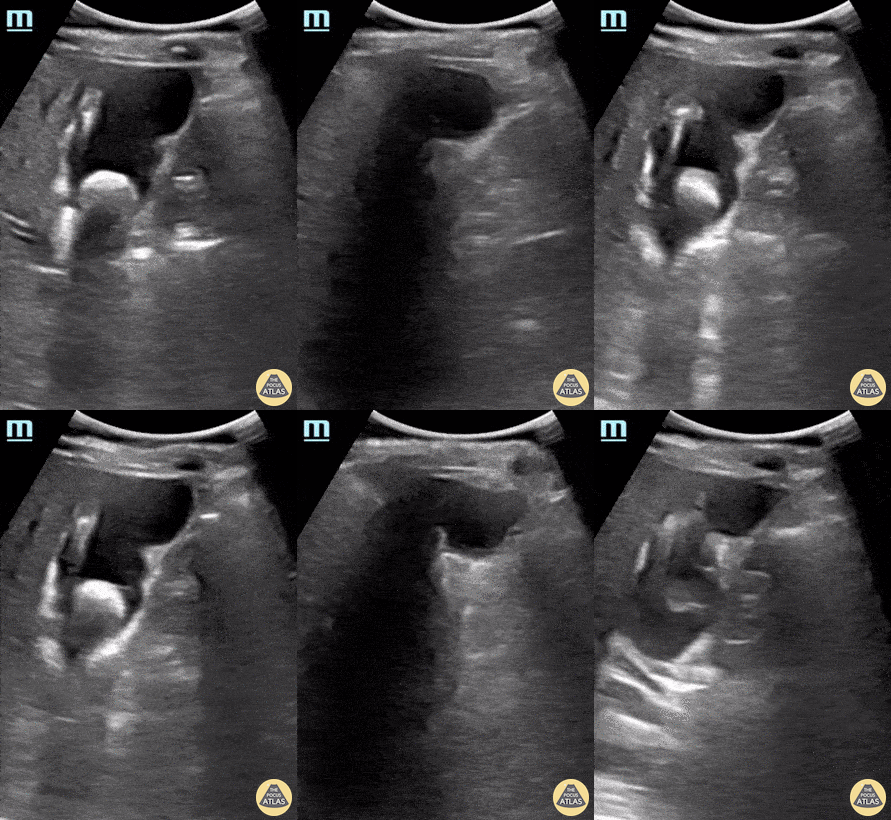 Cholelithiasis. Used with permission of the POCUS Atlas. Content License
Image Source: https://www.thepocusatlas.com/hepatobiliary/cholelithiasis
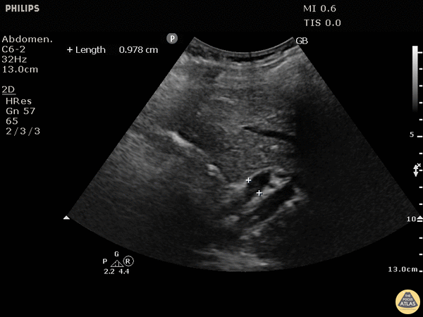 Dilated Common Bile Duct. Used with permission of the POCUS Atlas. Content License.
Image source: https://www.thepocusatlas.com/hepatobiliary/837oxd7ko4uk6awtz1l01zg3fpr2o9
Examiner Prompts (ABEM Domains and Rationale)
- Initial Reasoning: “What’s at the top of your differential, and what supports it?”
Ideal Response: Acute cholecystitis vs. ascending cholangitis. Supported by fever, RUQ pain, elevated bilirubin, and dilated CBD. (Synthesis) - Management Pathway: “What is your immediate management plan?”
Ideal Response: Start broad-spectrum IV antibiotics (eg, piperacillin-tazobactam), aggressive IV fluids, pain and nausea control, early surgical and GI consultation. (Management) - Clinical Turn: “Surgery is delayed, and the patient becomes hypotensive (BP 82/48) after fluids. What’s next?”
Ideal Response: Escalate to vasopressors, broaden coverage, and coordinate emergent ERCP or percutaneous cholecystostomy. (Management + Communication) - Procedural Judgment: “Would you consider bedside biliary drainage or cholecystostomy?”
Ideal Response: Yes, if no immediate IR or OR access and patient unstable. Ultrasound can guide safe decompression if operator and resources allow. (Synthesis + Data Gathering) - Antiplatelet Consideration: “How does her DAPT status affect your procedural and coordination plan?”
Ideal Response: Recognize elevated bleeding risk. Communicate early with cardiology and surgery for risk-benefit alignment. (Communication + Management)
Critical Actions (with ABEM Domains and Rationale)
- Identifies overlapping cholecystitis/cholangitis presentation (Synthesis) = Integrates ultrasound and labs under diagnostic uncertainty.
- Initiates sepsis management and broad antibiotics promptly (Management) = Executes early goal-directed care.
- Coordinates with GI, surgery, and cardiology regarding procedural timing and risk (Communication) = Demonstrates interdisciplinary collaboration.
- Anticipates need for escalation (ERCP, IR, ICU transfer) and documents plan (Follow-up / Management) = Shows foresight and structured closure.
Teaching Pearl:
Gallbladder ultrasound cases move beyond pattern recognition to judgment under competing priorities: balancing hemodynamic instability, comorbid risk, and procedural timing. That’s the reasoning depth ABEM expects and simulation can reproduce.
Integrating This into Training
We recommend embedding ABEM-style oral cases into residency and fellowship ultrasound curricula as part of longitudinal simulation design. Short, structured oral scenarios can be incorporated into resident and fellow education to assess not only image acquisition, but also cognitive integration, situational awareness, and team communication.
For faculty, these sessions double as real-time coaching laboratories. These sessions also provide opportunities to assess reasoning fidelity and feedback precision. Programs can scaffold complexity across the academic year, moving from straightforward image-based decisions to high-acuity, multi-system scenarios that test adaptability and leadership under stress.
Practical Model for Implementation
1. Residency Integration Model
Programs can embed brief ABEM-style oral scenarios into existing POCUS or simulation rotations as structured cognitive drills. For instance, interns might complete a seven-minute oral case weekly, adapted from current ultrasound simulations, with each scenario emphasizing a specific ABEM domain, such as Data Gathering in a trauma FAST or Synthesis in a biliary case. These cases can also be practiced by organizing mock oral board practice sessions with residents. This helps residents become comfortable with the format and practice ultrasound terminology, clinical integration, and technical skills.
Faculty can assess clarity of reasoning, prioritization, and communication flow using concise feedback rubrics rather than image-acquisition checklists. Over time, these sessions can serve as formative cognitive milestones, tracking growth in decision-making, adaptability, and composure across the first training year.
2. Fellowship and Faculty Development Model
For advanced learners and educators, ABEM-style oral frameworks can be integrated into complex multidisciplinary simulations, blending ultrasound decision-making with crisis resource management, procedural prioritization, and team leadership.
Fellows or faculty can alternate between examiner and candidate roles, debriefing on diagnostic framing, cognitive load management, and communication strategy. The focus shifts from individual performance to the meta-skills of teaching and assessment design, developing a shared vocabulary for feedback and evaluation across the department.
This model transforms ultrasound simulation into a dual-purpose exercise: refining high-stakes clinical reasoning while strengthening the instructional precision of those who teach it.
Tips for Converting a Case
- Choose concise, high-yield, image-driven simulations.3
- Identify 3-4 decision points that reveal reasoning under uncertainty.4
- Use prompts testing communication, prioritization, and adaptability.3,4
- Incorporate a few ultrasound clips requiring interpretation, not recall.5,6
- Keep case timing aligned with ABEM’s 7-10 minute ultrasound scenario format.7
Quick Checklist
- Identify a high-yield, image-driven case.
- Define three or four pivotal decision moments.
- Draft a succinct opening stem.
- Create examiner prompts with one purposeful follow-up each.
- Outline three to five critical actions mapped to ABEM domains.
- Pilot it with a colleague or senior resident and refine based on response quality.
Wrapping It Up
Transforming ultrasound simulations into ABEM-style oral scenarios elevates training from technical practice to structured cognitive rehearsal, directly aligning with ABEM’s certifying exam expectations in ultrasound, procedures, triage decision-making, and communication.3-8 The ACEP Ultrasound Simulation and Fellow Education subcommittees are currently working on compiling a compendium of cases for use by residencies and fellowships in training their learners and preparing them for clinical practice and the ABEM Certifying Exam. Appendix B includes an example of a case that can be found in the compendium.
Disclaimer: The cases in the appendix may be used for training and preparing learners, and have been developed by members of the fellow education subcommittee and therefore, may differ slightly than the final case format of the certifying exam.
Special thanks to the ACEP Ultrasound Section for fostering innovation in simulation-based learning and to the educators who continue to bridge hands-on ultrasound with the art of clinical judgment.
Citations
- Lewiss RE, Pearl M, Nomura JT, et al. CORD-AEUS consensus on emergency ultrasound education. Acad Emerg Med. 2013;20:1351-7.
- Soni NJ, Arntfield R, Kory P, et al. Multispecialty consensus on a POCUS basic skills checklist. Ultrasound J. 2022;14:1-15.
- Hilbert, Megan. “Hot Seat – with John Kendall for the US Component of Abem Certifying Exam.” Edited by Laura Oh, ACEP Emergency Ultrasound Section Newsletter. 5 May 2025, www.acep.org/emultrasound/newsroom/may-2025/hot-seat--with-john-kendall-for-the-us-component-of-abem-certifying-exam
- Unanyan, Mary. “Inside the New ABEM Certifying Exam: A Conversation on What’s Ahead.” Common Sense: Voice of the American Academy of Emergency Medicine. 2025;32(5):13-14,31-35.
- “Certifying Exam Content.” ABEM, www.abem.org/get-certified/certifying-exam/certifying-exam-content/. Accessed November 20, 2025.
- “Certifying Exam Scoring.” ABEM, www.abem.org/get-certified/certifying-exam/certifying-exam-scoring/.
- “Certifying Exam - Case Materials.” Certifying Exam Case Materials- Ultrasound, ABEM, December 2024. www.abem.org/wp-content/uploads/2024/12/Case-Materials_Ultrasound.pdf Accessed March 18, 2026.
- “Certifying Exam - Ultrasound List.” Certifying Exam Content, ABEM, May 2025, www.abem.org/wp-content/uploads/2025/04/Certifying-Exam_Procedures-List.pdf Accessed March 18, 2026.
Additional Resources
- ACEP Ultrasound SIM Case Library https://www.acep.org/ultrasound
- ABEM Certifying Exam Overview https://www.abem.org/public/become-certified/certifying-exam
- ACEP Emergency Ultrasound Section ultrasoundsection@acep.org
- The POCUS Atlas – Trauma Ultrasound Collection: https://www.thepocusatlas.com/bb-trauma
- The POCUS Atlas. Hepatobiliary Ultrasound Collection. Available at: https://www.thepocusatlas.com/hepatobiliary-1
- American College of Emergency Physicians. Emergency ultrasound guidelines. [policy ststement] Ann Emerg Med. 2017;69:e27- e54.
- Ilgen JS, Regehr G. Simulation for diagnostic reasoning: re-creating the matrix of medicine. Acad Med. 2019;94:1298-1304.
- Cook DA, Hamstra SJ, Brydges R, et al. Instructional design features in simulation-based education: a meta-analysis. Med Teach. 2013;35:e867- e898.
- American Board of Emergency Medicine. Certifying Exam Content Specifications. American Board of Emergency Medicine; 2023.
- Jang TB, Ruggeri W, Dyne PL. Learning the FAST exam: training fast and learning faster. Emerg Med Clin North Am. 2010;28:131-143.
- Blehar DJ, Barton B, Gaspari R. Learning curves in emergency ultrasound. Acad Emerg Med. 2015;22:574-582.
Appendix A: ABEM Domains Reference
|
Domain |
Core Focus |
Performance Indicators |
|
Data Gathering |
The ability to identify and obtain key information efficiently through focused history, targeted exam, and appropriate diagnostic testing, including point-of-care ultrasound. |
Strong performers collect data purposefully. They ask focused questions, order appropriate studies, and use bedside tools like ultrasound with intent and clinical relevance. |
|
Synthesis |
The ability to interpret findings, integrate information, and form a clear working diagnosis. |
Examiners look for organized reasoning: a concise, prioritized differential and a clear link between data, diagnosis, and decision-making. |
|
Management |
The ability to act decisively on your interpretation, prioritize interventions, and coordinate care under pressure. |
Effective candidates anticipate instability, initiate resuscitation early, involve consultants appropriately, and demonstrate structured prioritization in real time. |
|
Communication |
The ability to clearly convey your plan to patients, nurses, and consultants while maintaining composure and leadership presence. |
Examiners value calm, structured communication that directs the team, keeps everyone aligned, and shows empathy and professionalism even under stress. |
|
Appendix B: ULTRASOUND CANDIDATE TASK SHEET Case 1 ROOM # |
|
CASE PARAMETERS |
|
This is a 10-minute ultrasound case. During this case, you will acquire 2-3 ultrasound images and interpret 2-3 ultrasound videos. Please know that the examiner may interrupt you to move through the case in a timely manner. Expectations for Standardized Patient Interactions Upon entering the room, the examiner will introduce the patient. You should interact with the standardized patient as you would any patient in a clinical situation, but please:
Expectations for Examiner Interactions
Expectations for Ultrasound Machine Manipulation
|
|
PATIENT INFORMATION |
|
A 32-year-old male with no past medical history presents with chest pain and shortness of breath after a motorcycle accident. |
|
PHYSICAL EXAM FINDINGS |
|
Vital Signs: BP 89/57, HR 112, R 23, T 98.7°F (37.1°C), SpO2 89% |
|
TASK STATEMENT |
|
Your tasks are as follows:
|
CASE FORMAT
The candidate reads the above task sheet and enters the room when ready.
Examiner Introductory Statements
“Hello, Dr. (Candidate name). I am Dr. (Examiner name), welcome to ABEM General. I am going to be your examiner for this 10-minute ultrasound case.”
“Just a reminder, with the ultrasound machine, I am happy to adjust any machine settings such as gain, depth, modes, etc., probe selection, or help you position the bed or machine to your liking. During the exam, if you have acquired an image you like, you may ask me to freeze the screen. Before we get started, do you have any questions?”
Examiner then introduces the patient:
“Patient is a 32-year-old male with no past medical history presenting to the trauma bay of your emergency department for evaluation of chest pain and shortness of breath after a motorcycle accident. Patient is complaining of bilateral chest pain and shortness of breath with unstable vital signs as above.”
Patient may start to interact or make statements here regarding his chest pain and shortness of breath.
Image Acquisition and Task Completion
REMINDER: Candidates are to vocalize their thoughts as they scan the standardized patient
Obtain an ultrasound of the right lung in the second intercostal space.
Examiner
“Dr. (Candidate Name), what I would like you to show me is how you would you use an ultrasound to evaluate the right lung in the second intercostal space.
CANDIDATE SHOULD SELECT LINEAR PROBE AT THIS POINT
This will be an ultrasound of a normal lung. Candidate should identify the following:
- Soft tissue
- Rib
- Pleura
- Lung Sliding (normal)
- A-Lines (artifact)
- B-lines (artifact)
- OPTIONAL: Using M-mode to assess lung sliding. Do not prompt.
Interpret an ultrasound video of the right lung in the second intercostal space.
Examiner
“Dr. (Candidate Name), Now I would like to draw your attention to the screen (standardized video) and assume that the video you are currently seeing is the video you obtained. Using the stylus that is provided, could you point any significant anatomic abnormalities, artifacts, or important structures that would be important to you when evaluating this trauma patient.”
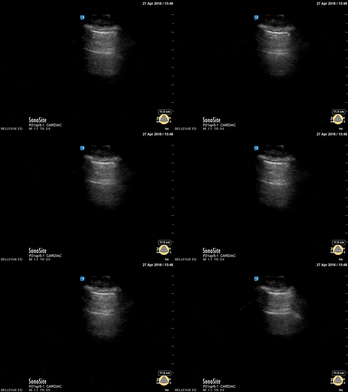 Video 1: Lung Sliding and A-lines. Used with permission of the POCUS Atlas. Content License. Image source: https://www.thepocusatlas.com/lung/skfn1nk408w1wvqph7pxsc5fr6pbs9
This is an ultrasound video of a normal lung. Candidate should identify the following:
- Soft tissue
- Rib
- Pleura
- Lung Sliding (normal)
- A-Lines (artifact)
Examiner
“The following ultrasound image is also obtained as part of your evaluation of the right lung.”
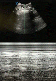 Image 1: Image reference: Adapted from The POCUS Atlas Pulmonary Collection. Used with permission of the POCUS Atlas. Content License.
This is an ultrasound video of a normal lung. Candidate should identify the following:
- Use of M-mode imaging modality
- Soft tissue
- Pleural Line
- Identify presence of sliding
- May mention “seashore sign”
Obtain an ultrasound of the right lung in the right upper quadrant.
Examiner
“Dr. (Candidate Name), what I would like you to show me is how you would you use an ultrasound to evaluate the right lung in the right upper quadrant.”
CANDIDATE SHOULD SWITCH TO CURVILINEAR PROBE AT THIS POINT
This will be an ultrasound of a normal lung. Candidate should identify the following:
- Soft tissue
- Rib
- Pleura
- Lung Sliding (normal)
- A-Lines (artifact)
- B-lines (artifact)
- OPTIONAL: Using M-mode to assess lung sliding. Do not prompt.
Interpret an ultrasound video of the right lung in the right upper quadrant.
Examiner
“Dr. (Candidate Name), Now I would like to draw your attention to the screen (standardized video) and assume that the video you are currently seeing is the video you obtained. Using the stylus that is provided, could you point any significant anatomic abnormalities, artifacts, or important structures that would be important to you when evaluating this trauma patient.”
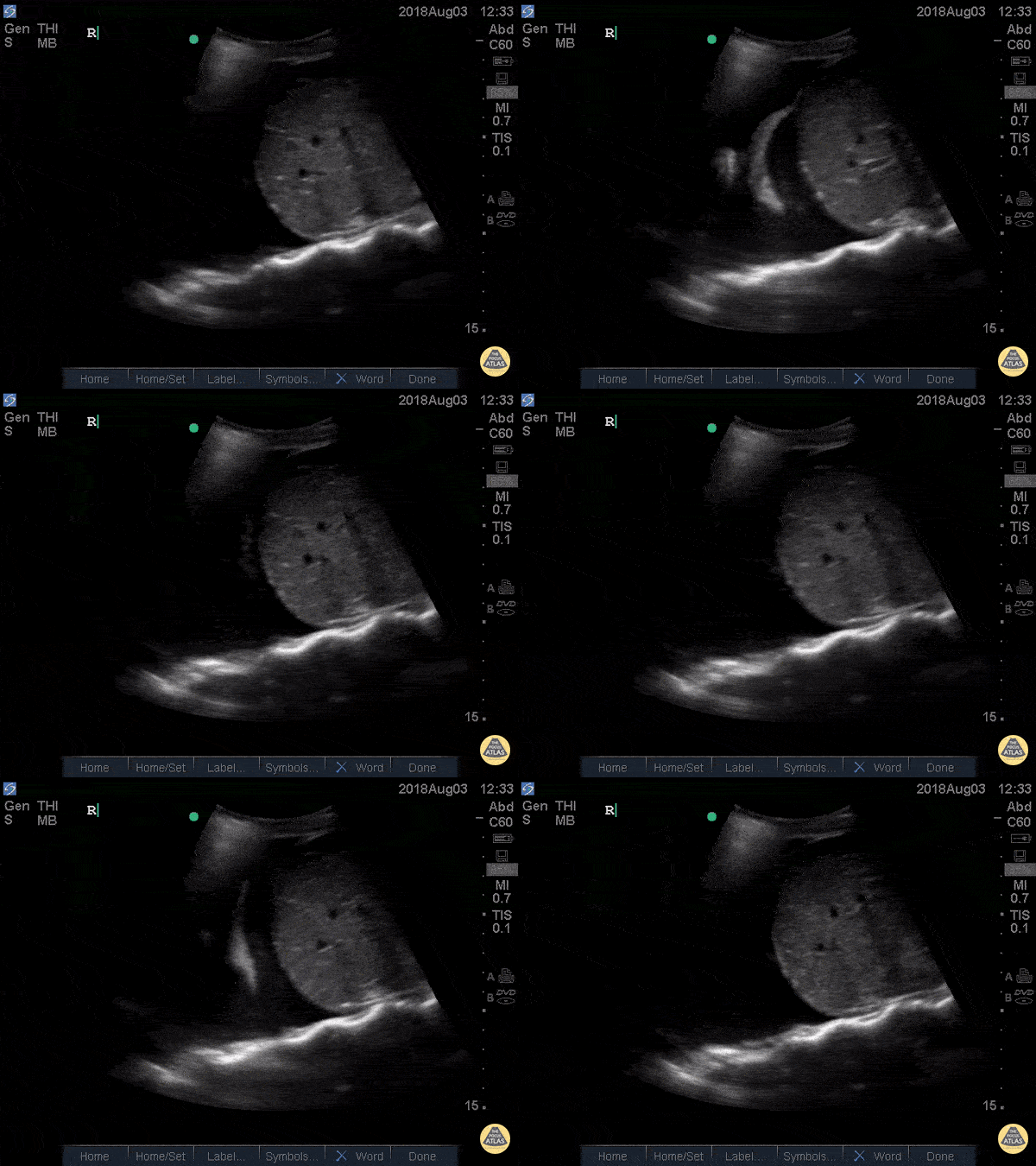 Video 2: Spine Sign. Used with permission of the POCUS Atlas. Content License. Image source: https://www.thepocusatlas.com/lung/ugrm87yr9owvaxeztp17ytwl3du4r0
This will be an abnormal ultrasound with pathology. Candidate should identify the following:
- Soft tissue
- Rib
- Pleura
- Liver
- Pleural Fluid Collection
- Lung floating in fluid collection
- Spine sign (Visualization of spinal in the posterior thoracic cavity)
Obtain an ultrasound of the left lung in the second intercostal space
Examiner
“Dr. (Candidate Name), what I would like you to show me is how you would use an ultrasound to evaluate the left lung in the second intercostal space.”
CANDIDATE SHOULD SELECT LINEAR PROBE AT THIS POINT
This will be an ultrasound of a normal lung. Candidate should identify the following:
- Soft tissue
- Rib
- Pleura
- Lung Sliding (normal)
- A-Lines (artifact)
- B-lines (artifact)
- Anatomic Consideration: Moving the ultrasound probe more lateral to avoid cardiac structures.
- M-Mode: If they did not use M-mode earlier when evaluating the right lung, prompt them to show you how to use it.
Interpret an ultrasound video of the left lung in second intercostal space
Examiner:
“Dr. (Candidate Name), Now I would like to draw your attention to the screen (standardized video) and assume that the video you are currently seeing is the video you obtained. Using the stylus that is provided, could you point any significant anatomic abnormalities, artifacts, or important structures that would be important to you when evaluating this trauma patient. You do not need to mention anything you have previously mentioned.”
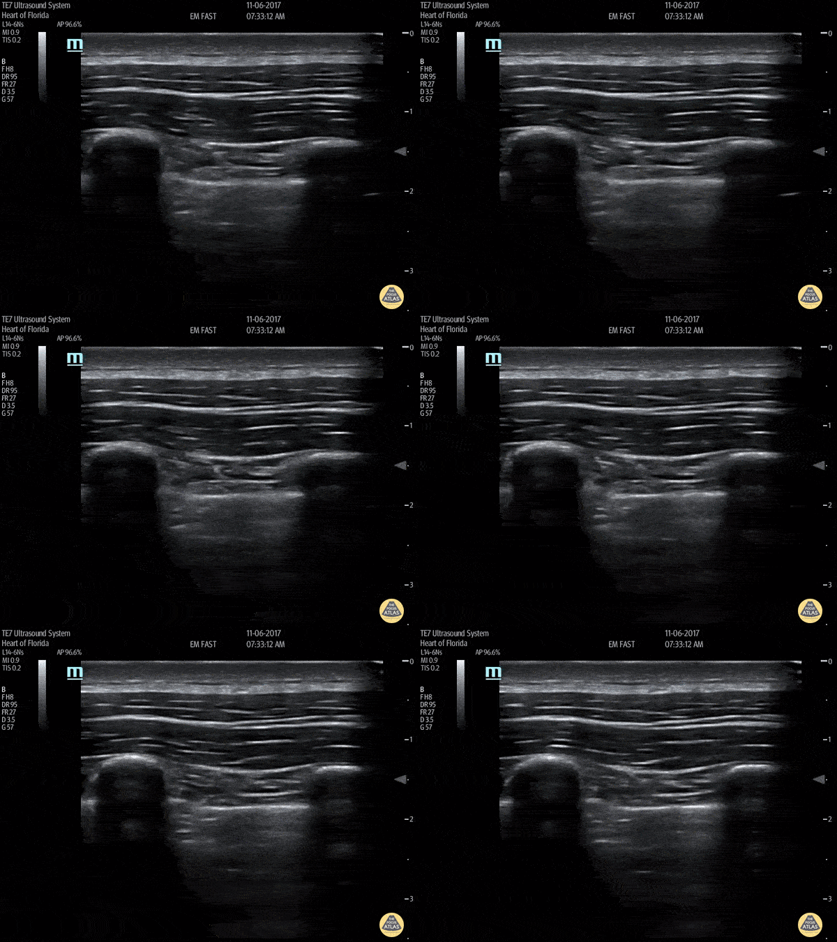 Video 3: No Lung Sliding. Used with permission of the POCUS Atlas. Content License.
Image source: https://www.thepocusatlas.com/lung/no-lung-sliding
This will be an abnormal ultrasound with pathology. Candidate should identify the following:
- Soft tissue
- Rib
- Pleura
- Loss of Lung Sliding (concerns for pneumothorax)
- Candidate may request M-mode or mention they would confirm their concerns with this modality.
Examiner:
If candidate does not request M-mode:
“Dr. (Candidate Name), is there anything else you would do with the ultrasound based on the above ultrasound video?
If candidate requests M-mode after prior video or once prompted:
“The following image is obtained given concerns for underlying pathology”
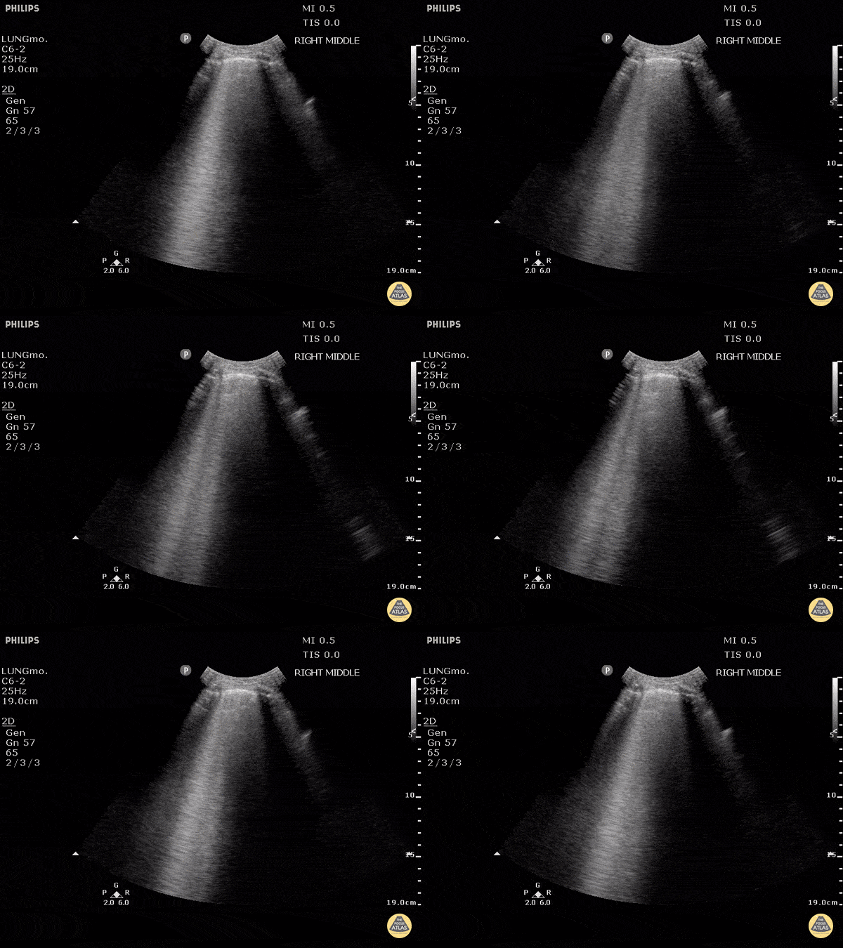
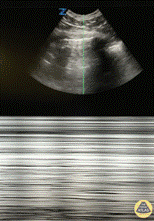 Image 2:Image reference: Adapted from The POCUS Atlas Pulmonary Collection.
Used with permission of the POCUS Atlas. Content License.
This will be an abnormal ultrasound with pathology. Candidate should identify the following:
- Soft tissue
- Rib
- Pleura
- Loss of Lung Sliding and concerns for pneumothorax
- May mention “stratosphere” or “barcode sign.”
Closing Statement
Examiner:
“Thank you Dr. (Candidate Name), that concludes your case.”
|
Appendix C: ULTRASOUND CANDIDATE TASK SHEET Case 2 ROOM # |
|
CASE PARAMETERS |
|
This is a 10-minute ultrasound case. During this case, you will acquire 2-3 ultrasound images and interpret 2-3 ultrasound videos. Please know that the examiner may interrupt you to move through the case in a timely manner. Expectations for Standardized Patient Interactions Upon entering the room, the examiner will introduce the patient. You should interact with the standardized patient as you would any patient in a clinical situation, but please:
Expectations for Examiner Interactions
Expectations for Ultrasound Machine Manipulation
|
|
PATIENT INFORMATION |
|
A 71-year-old female with past medical history of ESRD presents with chest pressure, shortness of breath, and abdominal distention after missing her last week of dialysis. |
|
PHYSICAL EXAM FINDINGS |
|
Vital Signs: BP 183/119, HR 103, R 22, T 98.4°F (36.9°C), SpO2 86% |
|
TASK STATEMENT |
|
Your tasks are as follows:
|
CASE FORMAT
The candidate reads the task sheet and enters the room when ready.
Examiner Introductory Statements
“Hello, Dr. (Candidate name). I am Dr. (Examiner name), welcome to ABEM General. I am going to be your examiner for this 10-minute ultrasound case.”
“Just a reminder, with the ultrasound machine, I am happy to adjust any machine settings such as gain, depth, modes, etc., probe selection, or help you position the bed or machine to your liking. During the exam, if you have acquired an image you like, you may ask me to freeze the screen. Before we get started, do you have any questions?”
Examiner then Introduces the patient:
“Your patient is a 71-year-old female with past medical history of ESRD presenting with chest pressure, shortness of breath, and abdominal distention after missing her last week of dialysis. Her vitals are BP 183/119, HR 103, R 22, T 98.4°F (36.9°C), SpO2 86%.
Patient may start to interact or make statements regarding symptoms.
Image Acquisition and Task Completion
REMINDER: Candidates are to vocalize their thoughts as they scan the standardized patient
1. Obtain a parasternal long view of the heart
Examiner:
“Dr. (Candidate Name), your patient is complaining of chest pressure and you would like to evaluate his cardiac function. What I would like you to show me is how you would you use an ultrasound to evaluate the heart using a parasternal long view.”
CANDIDATE SHOULD SELECT PHASED ARRAY AT THIS POINT
This will be an ultrasound of a normal heart. Candidate should identify the following:
- Right ventricle (and discuss right heart strain)
- Aortic Outflow Tract
- Left atrium
- Left ventricle (and discuss ejection fraction and wall motion)
- Descending thoracic aorta
- Pericardium (and discuss pericardial effusion)
2. Interpret an ultrasound video of the parasternal long view of the heart.
Examiner:
“Dr. (Candidate Name), Now I would like to draw your attention to the screen (standardized video) and assume that the video you are currently seeing is the video you obtained. Using the stylus that is provided, could you point any significant anatomic abnormalities, artifacts, or important structures that would be important to you when evaluating this trauma patient.”
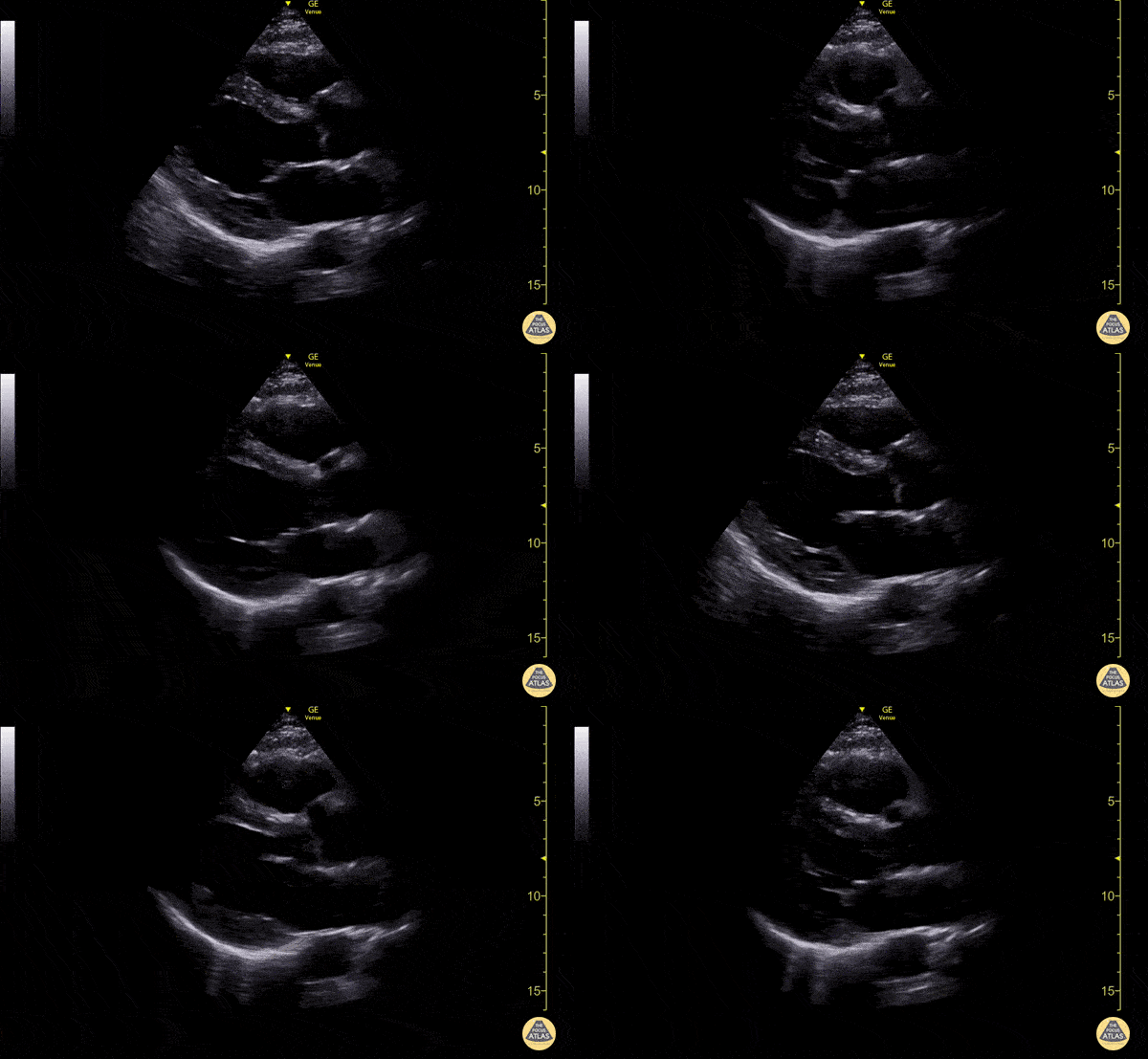 Video 1: Normal Parasternal Long Axis (PLAX) View. Used with permission of the POCUS Atlas. Content License.
Image source: https://www.thepocusatlas.com/normal-cardiac-anatomy/normal-parasternal-long-axis-plax-view
This is an ultrasound video of a normal PSLA. Candidate should identify the following:
- Right ventricle (lack of right heart strain)
- Aortic Outflow Tract
- Left atrium
- Left ventricle (normal ejection fraction)
- Descending thoracic aorta
- Pericardium (lack of pericardial effusion)
3. Obtain an ultrasound of the right lung at the second intercostal space.
Examiner:
“Dr. (Candidate Name), you suspect a component of fluid overload as part of this patient’s symptoms. What I would like you to show me is how you would you use an ultrasound to evaluate the right lung at the second intercostal space in regard to this patient’s shortness of breath.”
CANDIDATE SHOULD SELECT (CURVILINEAR OR PHASED ARRAY) AT THIS POINT
This will be an ultrasound of normal lung on a standardized patient. Candidate should identify the following:
- Soft tissue
- Rib (Artifact – posterior shadowing)
- Pleura
- Lung sliding (normal)
- A-lines (Artifact)
- B-Lines (Artifact)
4. Interpret an ultrasound video of the right lung at the second intercostal space.
Examiner:
“Dr. (Candidate Name), Now I would like to draw your attention to the screen (standardized video) and assume that the video you are currently seeing is the video you obtained. Using the stylus that is provided, could you point out any significant anatomy, anatomic abnormalities, artifacts, or measurements that would be important to you when evaluating this patient.”
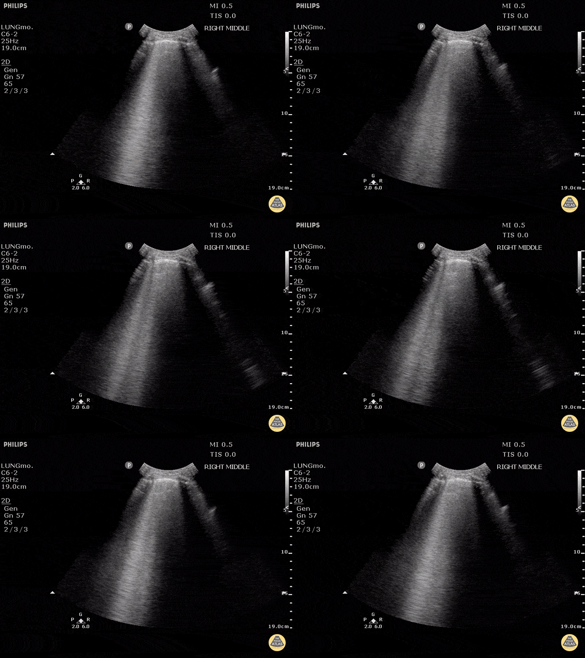 Video 2: B-Lines. Used with permission of the POCUS Atlas. Content License.
Image source: https://www.thepocusatlas.com/lung/q7shylclnfsc3w5qezhq9rmxee06vk
This will be an abnormal ultrasound with pathology. Candidate should identify the following:
- Soft tissue
- Rib (Artifact – posterior shadowing)
- Pleura
- Lung sliding (normal)
- B-Lines (Artifact present)
5. Obtain an ultrasound of the abdomen to assist in an ultrasound guided paracentesis.
Examiner:
“Dr. (Candidate Name), as you complete your exam you notice significant distention of the abdomen with a fluid wave. You feel this distention is exacerbating this patient’s dyspnea and discomfort. What I would like you to show me is how you would you use an ultrasound to obtain an image to assist in a therapeutic ultrasound guided paracentesis.”
CANDIDATE SHOULD SELECT (CURVILINEAR OR LINEAR) AT THIS POINT
This will be an ultrasound of normal abdomen on a standardized patient. Candidate should identify the following:
- Soft tissue
- Large bowel
- Small Bowel
6. Interpret an ultrasound video of the abdomen to assist in an ultrasound guided paracentesis.
Examiner:
“Dr. (Candidate Name), Now I would like to draw your attention to the screen (standardized video) and assume that the video you are currently seeing is the video you obtained. Using the stylus that is provided, could you point out any significant anatomy, anatomic abnormalities, artifacts, or measurements that would be important to you when setting up for this procedure.”
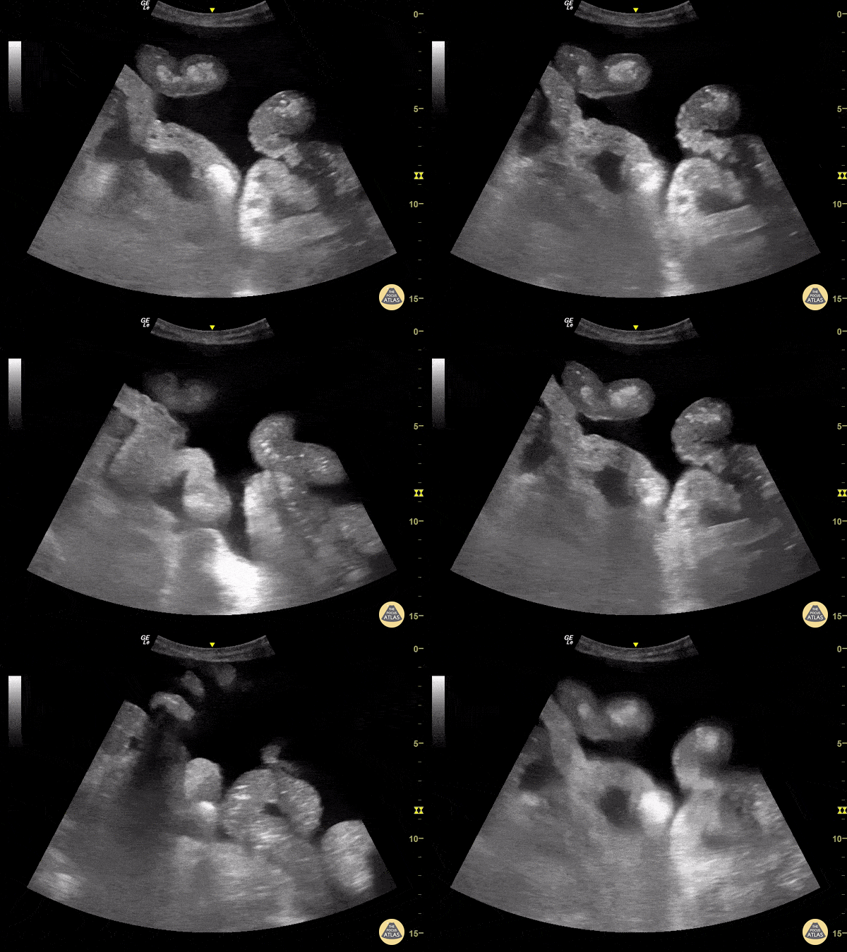 Video 3: Ascites. Used with permission of the POCUS Atlas. Content License.
Image source: https://www.thepocusatlas.com/bowel/ascites
This will be an abnormal ultrasound with pathology. Candidate should identify the following:
- Soft tissue
- Small Bowel
- Ascites
- Target for paracentesis (Avoidance of vasculature, largest fluid pocket, avoidance of organs)
Closing Statement
Examiner:
“Thank you Dr. (Candidate Name), that concludes your case.”