 American College of Emergency Physicians

Add-On Echo CPT Codes to Rev Up Your POCUS Reimbursement
ACEP Emergency Ultrasound Section Reimbursement and Coding Subcommittee
Dillon W. Casey, MD
Joshua Guttman, MD FRCPC, FAAEM, FACEP, FAEMUS
Christopher D Thom, MD, RDMS
Introduction
Current Procedural Terminology (CPT) codes are the 5-digit codes that identify specific procedures. Coders will assign these codes to the procedures we do during an ED visit and submit those codes to the payer. This communicates with the payer what service was performed and in turn drives reimbursement. CPT codes are updated annually by the American Medical Association. The vast majority of the codes do not change on an annual basis, but codes can be changed or new codes added in response to new technology or new procedures. Members of the ACEP Emergency Ultrasound Section Subcommittee on Coding and Reimbursement analyze the CPT codes and provide a comprehensive list of POCUS-related codes for the benefit of the section.
Within cardiac POCUS, the CPT code typically used is 93308, which is the code to denote a limited cardiac ultrasound. Since a basic cardiac POCUS uses only B-mode ultrasound to assess for pericardial effusion, systolic function, relative chamber size, and occasionally aortic root diameter, this code serves nicely. Cardiac POCUS traditionally did not include the use of color or spectral Doppler.
However, as cardiac POCUS has evolved and more advanced echocardiographic measures have been taken by more POCUS users - especially those with fellowship training in emergency medicine ultrasound, some of our cardiac POCUS exams go beyond the basics. Many of us are now using color Doppler for valvular regurgitation, pulsed-wave and tissue Doppler for diastolic function, and continuous wave Doppler to assess right atrial pressures and RV strain.
Using these additional Doppler modes adds complexity of our cardiac POCUS, but they do not meet criteria for a comprehensive echocardiogram (CPT 93306).
The AMA's CPT editorial panel understood this and therefore separate codes were created to reflect this reality.
Add-on Ultrasound Codes you Didn't know you Could use in POCUS
An add-on code is a CPT code that does not stand alone but is added on to a primary code when additional work is performed. These are denoted with a + symbol before the 5-digit code. Two of these codes are relevant to advanced cardiac POCUS and are added on to the primary 93308 code.
+93321 is for a “Doppler echocardiography pulse wave and/or continuous wave with spectral display, limited study”
+93325 is "Doppler echocardiography, color flow velocity mapping."
Case 1
A 72-year-old female presents for shortness of breath. She reports worsening dyspnea on exertion over the preceding 2 weeks, along with increased lower extremity edema bilaterally. On bedside ultrasound, the LVEF is normal, there is no pericardial effusion, and RV size and TAPSE measurement is normal, and the aortic root is normal in diameter. Upon careful inspection of the parasternal long images, the ED physician notes a subtle prolapse of the posterior leaflet of the mitral valve in diastole. Given this, color Doppler is applied and a severe, eccentric mitral regurgitation jet is noted.
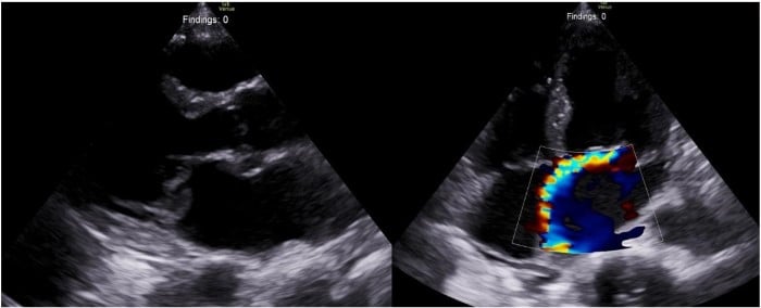
In your POCUS documentation, you include the medical necessity for the limited echo AND for using color Doppler (“color Doppler flow was utilized to assess for mitral regurgitation”). You document the limited echo findings AND the color Doppler flow findings (“color Doppler revealed severe, eccentric mitral regurgitation is present”). Lastly, you ensure a representative color Doppler image is archived along with your other echo images.
Your coder can now bill both CPT 93308 and the add-on 93325.
Case 2
A 66-year-old male presents for shortness of breath. He notes gradually worsening dyspnea on exertion over the preceding 4 months, along with increased weight gain and orthopnea. He has not had any chest pain or fever. On bedside ultrasound, the LVEF is normal, there is no pericardial effusion, and RV size and TAPSE measurement is normal, and the aortic root is normal in diameter. However, you do notice that the left atrium appears dilated and you decide to apply spectral pulsed-wave Doppler over the tip of the mitral valve leaflets in the apical four chamber to assess mitral inflow velocity. This reveals a restrictive filling pattern suggestive of diastolic dysfunction. You then confirm this via assessment of tissue Doppler e’ velocity and LA size measurement.
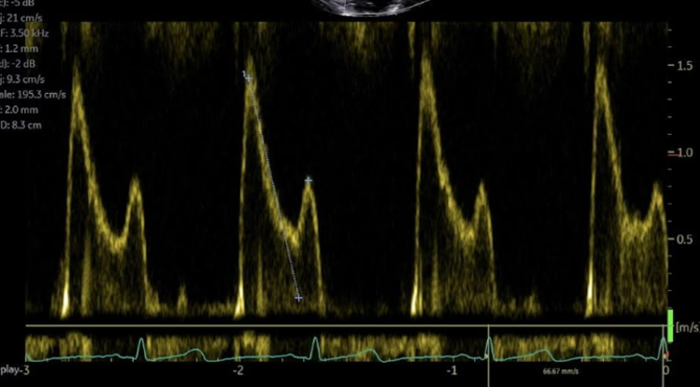
In your POCUS documentation, you include the medical necessity for the limited echo AND for using spectral Doppler (“spectral pulsed-wave Doppler was utilized for the assessment of diastolic function”). You document the limited echo findings AND the spectral Doppler findings (“Spectral pulsed-wave Doppler analysis of the mitral inflow velocities reveal an E wave of 1.5 m/s and an A wave of 0.7 m/s, with an E/A ratio of 2.1.”). Lastly, you ensure a representative color Doppler image is archived along with your other echo images.
Your coder can now bill both CPT 93308 and the add-on 93321.
wRVU’s associated with limited Echo and add-on CPT codes: