 American College of Emergency Physicians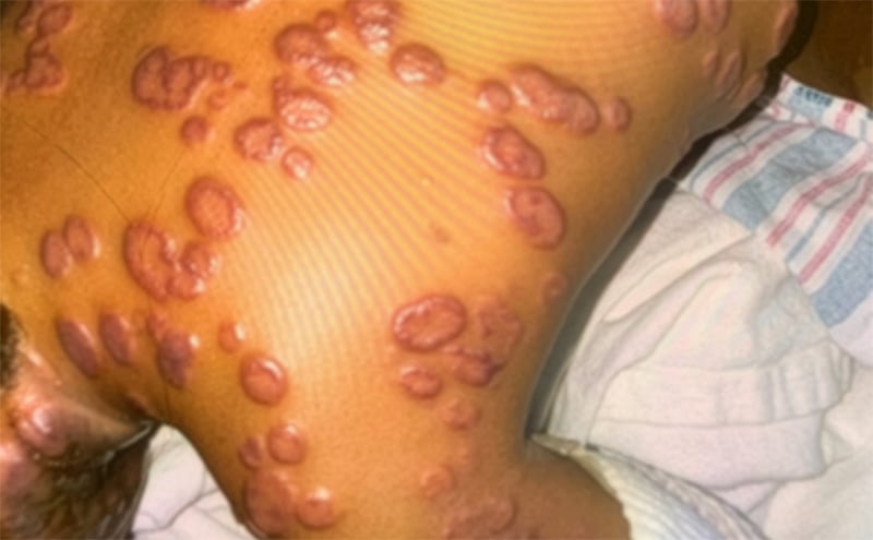

3rd Place EMage Winner: "Stop the bullae(ing):” Leukocyte Adhesion Deficiency Type 1 Presenting as Severe Bullous Pyoderma Gangrenosum
Sahar Sharif, MD
Andrea Frazier, MD
Ayesha Mirza, MD
Phoenix Children’s Hospital, Phoenix, AZ
Case Description
A 2-year-old otherwise healthy African American male presented to his primary care physician with ulcerative lesions on the scalp, neck, and buttocks with general malaise, runny nose, and nasal congestion. He was initially treated with outpatient oral antibiotics, but with worsening lesions, he was referred to the emergency department and then admitted to the general pediatric ward. Despite broad-spectrum antibiotic therapy, lesions continued to enlarge, and new ulcerating lesions developed throughout the body [Fig. 1], which were worse at the site of a recent central line placement [Fig. 2]. Additionally, white blood cell count increased as high as 78.85 K/mcL. Immunoglobulin and complement levels were within normal limits. Histopathological results showed exuberant diffuse dermal neutrophilic infiltrate with surrounding edema and granulation. Dermatologic findings were correctly identified as pyoderma gangrenosum (PG). Symptoms began to resolve with a regimen of prednisone, cyclosporine, intravenous immunoglobulin (IVIG), Tacrolimus, and Infliximab. The patient was found to have absent CD11/CD18 expression and a compound heterozygote for two variants in ITGB2 gene on genetic testing, confirming the underlying diagnosis of leukocyte adhesion deficiency (LAD).
Discussion
This case demonstrates a diagnostically challenging presentation of two rare pediatric conditions. Although LAD is considered in cases of delayed separation of the umbilical stump and severe invasive infections, it should also be considered in the differential diagnosis of abnormal wound healing and recurrent skin ulcers. LAD1 is associated with severe morbidity and early mortality, thus making a timely diagnosis essential. PG has a distinctive cutaneous ulceration with a purple edge. Half of PG cases are associated with a specific systemic disorder of rheumatologic, immunologic, or hematologic origin. The primary treatment of PG is high-dose corticosteroids and careful wound care.
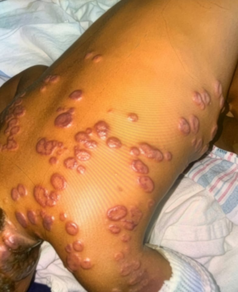
Figure 1. Numerous violaceous plaques and denuded bullae present on the trunk. Similar lesions were present on neck, bilateral inguinal folds, buttocks, and upper and lower extremities.
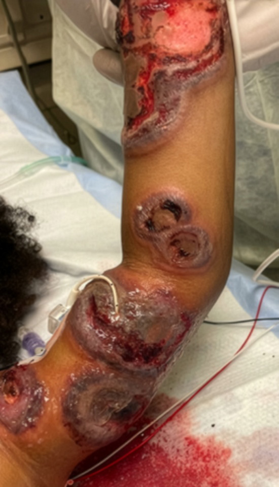
Figure 2. Lesions demonstrating pathergy phenomenon at site of PICC insertion. PICC, peripherally inserted central catheter.