 American College of Emergency Physicians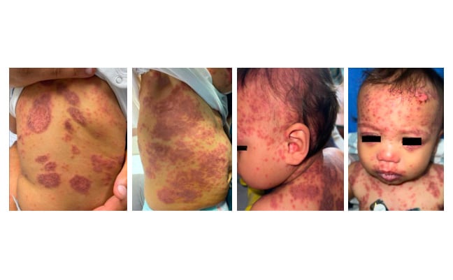

2nd Place EMage Winner: Maculopapular Lesions in a 12-Month-Old
Lisa Pace
University of Florida – Jacksonville, Wolfson Children’s Hospital
Case Presentation
A 12-month-old fully vaccinated male presented with a rash for 6 weeks. The rash initially presented with 5-6 circular lesions on his back, which were slightly raised, pink patches. The patient was given hydrocortisone cream by his pediatrician with no observed improvement. Three weeks later, the rash spread downwards towards his abdomen and legs. Then he was started on fluconazole and topical Nystatin for 2 weeks. One day before admission, the rash spread to his face. He was evaluated by an outside facility and diagnosed with hand-foot-mouth disease. Prescriptions for cephalexin and prednisone were provided, and he was discharged home. On the day of admission, his facial rash progressed with new-onset eye and mucosal involvement. There was no history of fever.
Physical examination was notable for diffuse, erythematous maculopapular lesions with crusting and tenderness throughout the scalp and face, extending down towards the back of the neck and ears, and anterior and posterior torso including buttocks and lower extremities, in addition to periorbital and conjunctival erythema and purulent eye discharge with lesions to the oral mucosa (Figures 1-4). The patient was admitted to the general pediatrics floor of our facility and was promptly transferred to the nearest burn center due to rapid progression of symptoms with sloughing of skin and mucosal membranes involving > 30% body surface area (Figures 5-10).
Laboratory studies were negative, including herpes simplex virus, varicella zoster virus, mycoplasma, adenovirus, and chlamydia pneumoniae. Ophthalmologic examination showed corneal involvement and conjunctival ulceration (Figure 11). A skin biopsy showed reactive infectious mucocutaneous eruption (RIME). Initial treatment regimen included moxifloxacin drops, cefepime, azithromycin, acyclovir, and intravenous immunoglobulin (IVIG). Later, he was transitioned to etanercept, IVIG, steroids, and cyclosporine. He was discharged home with cyclosporine and moxifloxacin drops for a 1-month duration. Due to diffuse healing of lesions (Figure 12), a final dressing change was completed prior to discharge home.
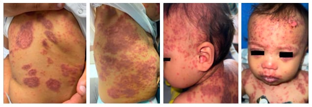
Figures 1-4: Photos of the back, right flank, lateral face, and scalp taken in the emergency department.
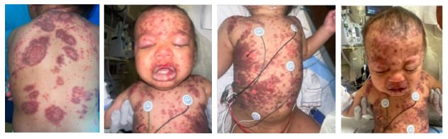
Figures 5-8: Photos of the face, back, and anterior torso taken in the general inpatient unit 4-5 hours after admission.
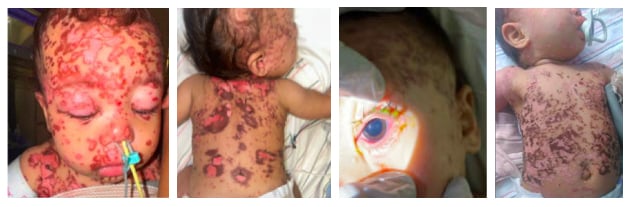
Figures 9-12: Photos taken at the burn center, including conjunctival involvement and improvement of rash.