Update on the Current PA and NP EUS Literature
ACEP Ultrasound Advanced Practice Providers Subcommittee
Increasing numbers of PAs and NPs are learning and incorporating Point-of-care ultrasound (POCUS) into their clinical practice. Along with this increased use has come additional contributions to the growing body of literature demonstrating that, with proper training, medical professionals at all levels have the ability to become proficient in EUS.
For the purposes of this review, we would like to present some of the recent data demonstrating PA and NP ability to obtain and interpret adequate bedside ultrasound imaging in a variety of modalities.
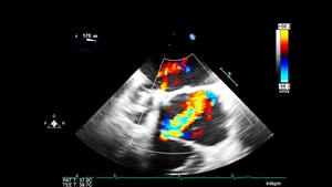
Focused Cardiac Ultrasound
A pilot study of physician assistant-focused cardiac point-of-care ultrasound assessment.1
Purpose: Determine if PAs can accurately obtain and interpret left ventricular function (LVF).
Methods: Eight Critical Care PAs obtained a total of 80 Focused Cardiac Ultrasounds (FoCUS) assessing LVF. Patents then underwent formal ECHO and results of the 2 independent studies were compared.
Results: Of the 80 scans obtained, PAs were 91% accurate when compared to findings on formal echocardiography. A total of 49 scans were considered abnormal by a PA. Of these scans, 45 were also considered abnormal by the cardiologists, with the 4 contradictory exams also being deemed normal by cardiology. A total of 31 scans were considered normal by a PA, and all but one was also considered normal by cardiology.
The sensitivity and specificity of PA performed FoCUS compared to formal ECHO for abnormal LVF was 0.91 (95% Cl, 0.79 to 0.97) and 0.96 (95% Cl, 0.81 to 0.99).
Conclusions: PAs with appropriate training are able to accurately assess left ventricular function using FoCUS.
For comparison, a study comparing emergency medicine resident performed FoCUS and formal echocardiography performed by a cardiologist found agreement in 91% of patients.2
FAST (Focused Assessment with Sonography in Trauma)
Advanced practice providers demonstrate proficiency in FAST after directed training.3
Purpose: Determine if PAs and NPs can accurately perform FAST exams after receiving a brief training that mirrors that given to physicians.
Methods: Eleven PAs and NPs underwent on-line and hands-on training on performing and interpreting FAST exams. Both pre- and post-tests were administered, and a 6-month post-test was administered to evaluate retention.
Results: 91% of the initial 17 scans performed by PAs and NPs after the training period were considered technically adequate when reviewed by emergency department US faculty. Due to lack of positive scans, evaluating image interpretation was limited.
Conclusions: PAs and NPs who completed a brief training, similar to that provided to physicians, are able to adequately perform FAST exams.
For comparison, a study of emergency physicians found them to be 97% accurate.4
Pneumothorax
Ultrasound detection of pneumothorax with minimally trained sonographers: a preliminary study.5
Purpose: Determine if non-physician providers can use ultrasound to accurately detect pneumothoraces.
Methods: Twenty-two PAs, U.S. Army medics, special ops forces, veterinary technicians, and food service inspectors who had no prior formal US training underwent a 10-minute lecture led by a PA. They then evaluated a total of 44 porcine thoraces for the absence or presence of pneumothorax.
Results: Twenty-one out of 22 pneumothoraces were successfully identified with 1 false-negative. All 22 normal hemithoraces were correctly identified. Sensitivity for pneumothorax was 95.5% (95% CI, 75-99%), specificity was 100% (95% CI, 81-100%).
Conclusions: With minimal training, non-physician providers can accurately identify pneumothoraces using ultrasound.
For comparison, a study evaluating accuracy of ED physician-performed US after a 2-hour course found a sensitivity of 86.4% and a specificity of 100% for detecting traumatic pneumothorax.6 Another study found that senior residents and attendings who were experienced with US had a 95% sensitivity and 99% specificity for detecting pneumothorax.7
Soft Tissue Abscesses
Ultrasound Detection of Soft Tissue Abscesses Performed by Non-Physician U.S. Army Medical Providers Naïve to Diagnostic Sonography.8
Purpose: Determine whether U.S. Army non-physician providers can use ultrasound to identify abscesses after minimal training
Methods: Thirty-one PAs and medics with no to minimal ultrasound training underwent a 30-minute didactic training followed by hands-on training for both clinical exam and ultrasound detection of soft tissue abscesses. They then evaluated 10 chicken breast models for presence of an abscess. Results were obtained separately for clinical exam and ultrasound findings. Nine participants were lost due to military operational requirements.
Results: Clinical examination was 73.5% sensitive (95% CI, 65.3-80.3%) for detecting abscesses and 77.2% specific (95% CI, 67.4-84.9%) with a diagnostic accuracy of 75% (CI 95%, 68.9-80.3%). Ultrasound was 99.2% sensitive (95% CI, 95.4-99.9%) and 95.5% specific (CI 95%, 88.5-98.6%) with a diagnostic accuracy of 97.7% (CI 95%, 94.6-99.2%). When compared with clinical exam alone, ultrasound changed the treatment plan in 25.4% of cases, and 94.6% of the time changed the treatment plan to the correct diagnosis.
Conclusions: With minimal training, PAs and medics can use ultrasound to identify abscesses more accurately than when utilizing the physical exam alone.
For comparison, study authors compared their results to those from similar studies in novice and expert physicians; in these studies, sensitivity for detecting abscess with ultrasound was 97.0-98.0%.
Soft Tissue Foreign Bodies
Detection of soft tissue foreign bodies by nurse practitioner-performed ultrasound.9
Purpose: Evaluate the accuracy of NP-performed US for detecting soft tissue foreign bodies
Methods: Ten NPs with no prior US experience underwent a 2-hour training that included both didactic and hands-on instruction. Their ability to identify wood, metal, and plastic foreign bodies in chicken thigh models was then assessed. Models were also scanned by experienced emergency physicians (EP) for comparison.
Results: Novice NPs had a sensitivity of 78.3% (CI 95% 66.4-86.9%) and specificity of 50% (CI 95% 29.9-70.1%). Experienced EPs had a sensitivity of 83.3% (CI 95% 55.2-95.3%) and specificity of 75% (CI 95% 30.1-95.4%). Sensitivity for identifying plastic foreign bodies was significantly worse in both groups (50% in both NP and EPs).
Conclusions: The authors concluded that their results fell in line with prior physician studies and that NPs can use US for detection of soft tissue foreign bodies after a brief training session.
Aorta
Point of Care Ultrasound Assessment of the Abdominal Aorta by Physician Assistant Students: A Pilot Study.10
Purpose: Evaluate the ability of PA students to acquire adequate ultrasound images of the abdominal aorta (AA).
Methods: Twenty PA students from 7 different programs reviewed online material and completed 4 precepted AA scans on 4 different mock patients. They were then asked to scan a fifth patient and their images were recorded and reviewed by 3 different ultrasound fellowship trained emergency medicine physicians for proper anatomical identification, aortic measurement, and interpretability of images.
Results: Ninety-five percent of PA students were able to obtain interpretable images of the distal aorta and longitudinal views of the aorta and 65% were able to obtain interpretable images of the proximal and mid-aorta.
Conclusions: After limited training, PA students were able to obtain adequate AA images at comparable levels as medical students and EM residents.
For comparison, 2 prior studies found that 87% of medical students were able to find interpretable images of the aorta after 9 hours of training, and that senior EM residents who had completed a 2-week US rotation and had extensive US experience (>150 scans) scored an 88% on image acquisition for abdominal aortic views.11,12 It is worth noting that the residents in the Schmidt study had more difficulty imaging the SMA and celiac arteries, suggesting that the lower scores PA students obtained of proximal and mid-aorta views may be reflective of an overall greater difficulty in imaging these areas.12
Conclusion
While this is not a comprehensive review, recent data continues to demonstrate that PAs and NPs could accurately utilize EUS ultrasound for a variety of indications. Further, it is likely that their ability and accuracy will continue to improve with experience.
In general, these studies enrolled low numbers of participants and most had limited time to train and evaluate participants. Thus, results may be difficult to compare to residency-trained providers or providers who have years of practice with EUS. Nevertheless, PAs and NPs in these studies consistently demonstrated the ability to accurately utilize EUS.
To date, there is no widely agreed upon consensus as to what constitutes baseline competency for either physician, PA, or NP performed EUS, which does limit interpretation of current data. Additional research should be done to establish baseline competency goals and further evaluate long-term skill and knowledge retention.
References
- Beaten R, Masinelli S. A pilot study for physician assistant-focused cardiac point-of-care ultrasound assessment. Chest. 2018;154(4):1121A.
- Farsi D, Hajsadeghi S, Hajighanbari MJ, et al. Focused cardiac ultrasound (FOCUS) by emergency medicine residents in patients with suspected cardiovascular diseases. J Ultrasound. 2017;20(2):133-8.
- Davis VW, Wallace JM, Ahern MT, et al. Mid-level providers demonstrate proficiency in FAST after directed training. Crit Ultrasound J. 2011; 3(2):111-3.
- Tsui CL, Fung HT, Chung KL, et al. Focused abdominal sonography for trauma in the emergency department for blunt abdominal trauma. Int J Emerg Med. 2008;1:183-7.
- Monti JD, Younggren B, Blankenship R. Ultrasound detection of pneumothorax with minimally trained sonographers: a preliminary study. J Sp Op Med. 2009;9(1):43-6.
- Abbasi S, Farsi D, Hafezimoghadam P, et al. Accuracy of emergency physician-performed ultrasound in detecting traumatic pneumothorax after a 2-h training course. Eur J Emerg Med. 2013;20(3):173-7.
- Nandipati KC, Allamaneni S, Kakarla R, et al. Extended focused assessment with sonography for trauma (EFAST) in the diagnosis of pneumothorax: experience at a community-based level I trauma center. Injury. 2011;42(5):511-4.
- LaDuke M, Monti J, Cronin A, et al. Ultrasound detection of soft tissue abscesses performed by non-physician U.S. Army medical providers naïve to diagnostic sonography. Mil Med. 2017;182(3):e1825-e1830.
- Atkinson P, Madan R, Kendall R. Detection of soft tissue foreign bodies by nurse practitioner-performed ultrasound. Crit Ultrasound J. 2014;6(1):2.
- Jaynstein DL, Baeten R, Bafuma P, et al. Point of care ultrasound assessment of the abdominal aorta by physician assistant students: A pilot study. JPAEA. Accepted for publication February 2020.
- Andersen GN, Viset A, Christian MO, et al. Feasibility and accuracy of point-of-care pocket-size ultrasonography performed by medical students. BMC Med Educ. 2014 Jul 28; 14:156.
- Schmidt JN, Kendall J, Smalley C. Competency assessment in senior emergency medicine residents for core ultrasound skills. West J Emerg Med. 2015 Nov; 16(6): 923–926.
Dayna Jaynstein, MSPAS, PA-C
Assistant Director of the Red Rocks PA Program in Arvada, CO
@jaynstein
Aaron Inouye, PA-C
Director at Large for the Society for Point of Care Ultrasound
@PAintheED