 American College of Emergency Physicians

POCUS Before CT: Delayed Sigmoid Perforation After Colonoscopic Polypectomy
Camden Fuller MD MPH
Siddharth Shanbhag BS
Theresa Kim MD
Keywords: Point-of-care ultrasound (POCUS); bedside ultrasound; emergency medicine; colonoscopy perforation; post-polypectomy perforation; feculent peritonitis; pneumoperitoneum; viscus perforation; septic shock
Abstract
Case Presentation
A 75-year-old woman presented to the emergency department with abdominal pain, hypoxia, and hypotension. Due to clinical instability, POCUS was used to identify pneumoperitoneum prior to advanced imaging, following a colonoscopy six days earlier.
Discussion
Viscus perforation is a serious, potentially fatal complication that requires prompt recognition by emergency physicians. While Computed Tomography (CT) imaging is the gold standard for diagnosis of pneumoperitoneum and viscus perforation, critically ill patients may not tolerate the time or transport required for advanced imaging. POCUS overcomes these challenges by providing real-time imaging at the bedside without interrupting resuscitation.
Case Presentation
A 75-year-old woman with hypertension, hyperlipidemia, atrial fibrillation on anticoagulation, and peripheral arterial disease with stenting and bilateral above-knee amputations presented to the emergency department with one day of hypoxia, hypotension, mottling, and abdominal pain with peritoneal signs. Notably, she underwent a colonoscopy with 3.5 cm sigmoid polypectomy six days prior. On presentation, she met criteria for septic shock with peritoneal signs on exam. She was resuscitated with antibiotics, intravenous fluids, vasopressors, and endotracheal intubation, and required cardioversion for ventricular tachycardia. CT imaging was delayed by ongoing bedside resuscitation and an episode of ventricular tachycardia that required emergent cardioversion in the scanner. A RUSH exam (Rapid Ultrasound in Shock and Hypotension) was performed to evaluate the etiology of her shock. POCUS demonstrated intra-abdominal free fluid, to-and-fro peristalsis, and a dilated bowel loop measuring 2 cm in diameter. (Figure 1, Video 1). Although ‘to and fro’ peristalsis is most often seen in patients with an intestinal obstruction, the bowel diameter of 2 cm did not support the diagnosis of a small bowel obstruction (SBO). There was concern for paralytic ileus in the setting of pneumoperitoneum.
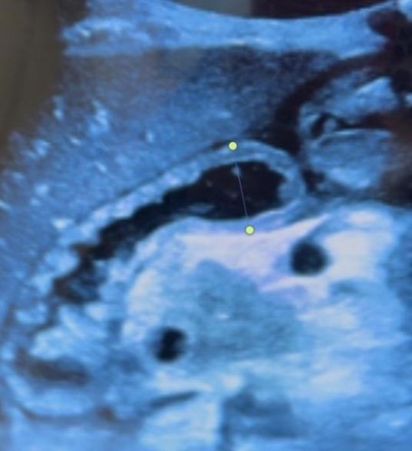
Figure 1. POCUS demonstrating a dilated bowel loop with intraluminal feculent material and extraluminal free fluid. Bowel diameter measures 2 cm.
Video 1. Abdominal POCUS demonstrating to-and-fro peristalsis, an abnormal motility pattern most commonly associated with intestinal obstruction.
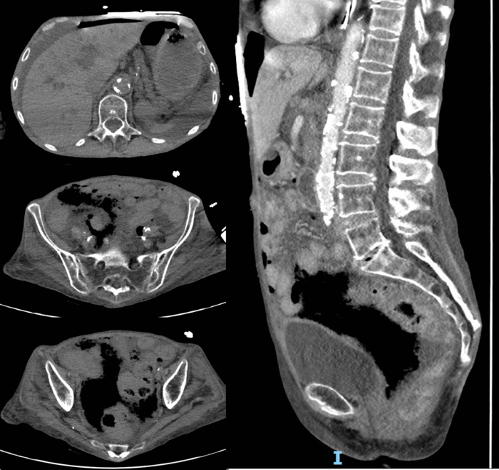 Figure 2. CT Abdomen and Pelvis image showing extensive pneumoperitoneum and free abdominal fluid secondary to perforated viscus
Discussion
Colonoscopy is one of the most commonly performed procedures in the United States, with indications including colorectal cancer screening, diagnosis, and therapeutic intervention. Although rare, viscus perforation is among the most serious complications, as delayed recognition can rapidly progress to feculent peritonitis and pneumoperitoneum. Current literature notes that perforation rates following colonoscopies are estimated to range between 6.6 to 9 per 10,000 colonoscopies; colonoscopy with polypectomy was noted to increase the risk of perforation, with delayed recognition and physical signs as factors associated with poor outcomes.1-4 While CT imaging is considered the gold standard for diagnosing pneumoperitoneum, critically unstable patients may not tolerate transportation to the scanner or delays associated with obtaining advanced imaging.
As viscus perforation is a serious and potentially fatal complication, prompt recognition is essential for emergency medicine physicians. Despite the low frequency of perforation rates, the large number of colonoscopies performed annually in the United States — estimated to exceed 15 million in 20255 - makes it likely that emergency medicine physicians will encounter post-colonoscopy perforation patients during their careers. While rare, colonoscopy carries a risk of perforation that increases with age, sex, polypectomy of > 2.0 cm, and vascular co-morbidities.6 Perforation most frequently occurs at the sigmoid colon, and emerging data suggest a potential association between colonoscopy and colonic ischemia.7-8
This patient had multiple risk factors for perforation, including advanced age, low BMI, significant vasculopathy, and resection of a 3.5 cm polyp during colonoscopy. The delay between colonoscopy and symptom onset suggests these factors may have increased perforation risk while also impairing healing at the perforation site due to ischemia, causing delayed perforation.
Initial POCUS findings of free fluid and to-and-fro peristalsis raised concern for small bowel obstruction (SBO). However, the additional POCUS finding of a bowel diameter of 2 cm, along with the patient’s presentation and history, was not consistent with SBO. Prior studies have demonstrated that SBO typically presents with bowel dilation greater than 2.5cm, which is one of the most sensitive findings on POCUS used to rule in SBO as a differential diagnosis.9-12 Instead, our findings were suggestive of perforation-induced paralytic ileus.13
We highlight the importance of POCUS in expediting operative management in a critically ill patient who was actively decompensating. By identifying free fluid at the bedside in a patient in septic shock, we were able to expedite surgical consultation. Although the patient ultimately died, operative intervention offered her the only realistic chance of survival.
POCUS findings can be variable and are highly dependent on patient factors, variations in anatomy, and operator experience. Even experienced users can have difficulty obtaining adequate POCUS images if patients have an unfavorable body habitus, anatomical variants, or limitations in positioning. Though POCUS cannot definitively diagnose bowel perforation, in this patient’s case, it enabled us to rapidly infer that a perforated viscus was the most likely diagnosis. This case highlights the use of POCUS in a critically unstable patient for whom ongoing resuscitation delayed CT imaging, yet management was expedited through POCUS findings.
References
- Huffstetler AN, Fraiman J, Brownlee S, Stoto MA, Lin KW. An Estimate of Severe Harms Due to Screening Colonoscopy: A Systematic Review. J Am Board Fam Med. 2023;36(3):493-500. doi:10.3122/jabfm.2022.220320R2
- Levin TR, Zhao W, Conell C, et al. Complications of Colonoscopy in an Integrated Health Care Delivery System. Ann Intern Med. 2006;145(12):880. doi:10.7326/0003-4819-145-12-200612190-00004
- Reumkens A, Rondagh EJA, Bakker MC, Winkens B, Masclee AAM, Sanduleanu S. Post-Colonoscopy Complications: A Systematic Review, Time Trends, and Meta-Analysis of Population-Based Studies. Am J Gastroenterol. 2016;111(8):1092-1101. doi:10.1038/ajg.2016.234
- Lv XH, Lu Q, Wang ZJ, et al. Colonoscopy-Related Adverse Events in the 21st Century: An Updated Systematic Review and Meta-Analysis. Am J Gastroenterol. 2026;121(1):212-221. doi:10.14309/ajg.0000000000003429
- Roth A, Lazris A, Haskell H, James J. Overuse of Colorectal Cancer Screening and Surveillance. Am Fam Physician. 2025;112(4):452-455. https://www.aafp.org/afp/2025/1000/lown-right-care-colorectal-cancer-screening
- Lohsiriwat V. Colonoscopic perforation: Incidence, risk factors, management and outcome. World J Gastroenterol. 2010;16(4):425. doi:10.3748/wjg.v16.i4.425
- de’Angelis N, Di Saverio S, Chiara O, et al. 2017 WSES guidelines for the management of iatrogenic colonoscopy perforation. World J Emerg Surg. 2018;13:5. doi:10.1186/s13017-018-0162-9.
- Sadalla S, Lisotti A, Fuccio L, Fusaroli P. Colonoscopy-related colonic ischemia. World J Gastroenterol. 2021;27(42):7299-7310. doi:10.3748/wjg.v27.i42.7299
- Catena F, De Simone B, Coccolini F, Di Saverio S, Sartelli M, Ansaloni L. Bowel obstruction: a Narrative Review for All Physicians. World J Emerg Surg. 2019;14(1). doi:10.1186/s13017-019-0240-7
- Tamburrini S, Serra N, Lugarà M, et al. Ultrasound Signs in the Diagnosis and Staging of Small Bowel Obstruction. Diagnostics. 2020;10(5):277-277. doi:10.3390/diagnostics10050277
- Rosano N, Gallo L, Mercogliano G, et al. Ultrasound of Small Bowel Obstruction: A Pictorial Review. Diagnostics. 2021;11(4). doi:10.3390/diagnostics11040617
- Jang TB, Schindler D, Kaji AH. Bedside ultrasonography for the detection of small bowel obstruction in the emergency department. Emerg Med J. 2010;28(8):676-678. doi:10.1136/emj.2010.095729
- Langell JT, Mulvihill SJ. Gastrointestinal Perforation and the Acute Abdomen. Med Clin North Am. 2008;92(3):599-625. doi:10.1016/j.mcna.2007.12.004